La tétracycline, connue sous le nom commercial Sumycin, agit en bloquant la fixation de l’ARNt sur la sous-unité 30S ribosomale, interrompant l’élongation de la chaîne protéique bactérienne. Ce mécanisme confère une activité sur un spectre large, incluant bactéries Gram positives, Gram négatives, rickettsies et spirochètes. Sa biodisponibilité digestive varie selon la prise alimentaire et les interactions avec les ions divalents comme calcium et magnésium. Sa diffusion tissulaire est importante, notamment dans les voies respiratoires et génito-urinaires. L’élimination se fait par voie rénale et biliaire. Les effets indésirables incluent photosensibilisation, troubles digestifs et coloration dentaire en cas d’administration précoce. Les guides thérapeutiques mentionnent sumycin prix, en soulignant la nécessité de restreindre son utilisation afin de limiter les résistances acquises.
Loomaarst.ee
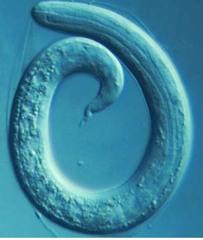
Definition of Tracheal Worms (Oslerus osleri) Oslerus osleri (formerly named Filaroides osleri) are parasites that infect the respiratory tracts of many mammals, causing a localized irritating and inflammatory reaction. They have been located in South Africa, New Zealand, Great Britain, France, India, Australia and the United States, and probably in other places as well. Oslerus osleri have been identified in mink, polecats, squirrel monkeys, cats, dogs and a number of Australian marsupials, among other species. While they are sometimes referred to as “lung worms,” Oslerus osleri actually infect the lumen and lining of their host animal’s trachea and larger bronchi. The lumen is the central cavity or hollow channel on the inside of a tube or tubular organ. The trachea is the tube-like organ that connects the mouth, nose and throat to the lungs; it is commonly called the “windpipe.” Bronchi are the large passageways that transport air from the trachea to and through the lungs. Tracheal worms are fairly small. They usually ranging from about 5 to 15 millimeters in length. The females tend to be larger than the males.
Causes of Tracheal Worms (Oslerus osleri) Tracheal worms do not require an intermediate host to complete their life cycle. In other words, they live most if not all of their lives inside of their canine hosts. This is called a “direct lifestyle.” Dogs become infected with Oslerus osleri through several routes. Puppies can become infected with tracheal worm larvae from the saliva of their mother, while she is licking or cleaning them. Dogs also can become infected by eating regurgitated food and by ingesting larvae from infected feces. Trachael worm larvae can be transferred through airway secretions, as well.
Oslerus osleri larvae enter the dog’s small intestine. They molt and migrate through the bloodstream into the trachea and bronchi, where they mature into adults. The worms trigger an inflammatory reaction inside the dog’s upper respiratory tract, which causes fibrotic nodules (lumps) to form inside the trachea. Eggs laid by adult tracheal worms hatch into larvae inside these thin-walled nodules and can be quite irritating. They are coughed up and swallowed by the infected dog, where they again land in the small intestine. The larvae are then either excreted in feces or migrate back to the trachea and large bronchi.
Symptoms of Tracheal Worms (Oslerus osleri) Despite their small size, tracheal worms can cause fairly severe illness in some companion dogs, while in others the signs are mild and nonprogressive. Dogs with tracheal worms may develop one or more of the following symptoms:
Cough (usually persistent/chronic, dry/unproductive and
Difficulty breathing (dyspnea; respiratory distress)
Wheezing sounds when breathing in (on inspiration); mild to
Panting (usually not pronounced except in advanced cases) Exercise intolerance
Retching (may be productive; may bring up white or blood-
Skin inflammation (dermatitis; rash; uncommon)
Dogs At Increased Risk Individual dogs can be infected with tracheal worms, but more commonly Oslerus osleri infection is a kennel-wide problem. This reportedly is especially true in large groups of greyhounds, although the reason for this particular association is not clear. Tracheal worms are seen mainly in young dogs less than 2 years of age. When they infect older dogs, these parasites often cause no significant symptoms.
Diagnosing Tracheal Worms (Oslerus osleri) In most cases, tracheal worms in dogs go undetected. When signs do show up, the diagnosis often can be made by finding the parasite eggs or larvae in fresh fecal samples. Unfortunately, Oslerus osleri larvae are often sluggish and only periodically shed in an infected dog’s stool. This makes bronchoscopy a better diagnostic technique.
Bronchoscopy involves inserting an endoscope, which is a wand-like medical instrument with a camera on its tip, down into the dog’s trachea. The camera enables the veterinarian to see the small, thin-walled, cream-colored growths that contain the parasites and protrude from the lining of the trachea. Sometimes, larvae can be seen peeking out from those nodules. The veterinarian can brush the nodules and examine the brushings under a microscope, either in a saline solution or in a special stain, to identify tracheal worm larvae. If this is not diagnostic, she can also use the endoscope to snip out and retrieve (biopsy) a small sample of affected tracheal tissue for submission to a diagnostic laboratory.
Chest X-rays (thoracic radiographs) can sometimes be helpful, especially if the infection is advanced and the nodules caused by the parasites inside the trachea are large and obvious. Finally, the lining of the trachea can be washed with a special solution, which is then drawn back into a syringe or tube and analyzed microscopically for the
presence of tracheal worm larvae. This procedure is known as a trans-tracheal wash.
Treating Tracheal Worms (Oslerus osleri) Treatment protocols for tracheal worms in companion dogs are still somewhat experimental. Some of the medications that have been used include albendazole, fenbendazole, levamisole, thiabendazole, thiacetarsamide sodium, diethylcarbamazine, prednisone and ivermectin. Some of these have been used successfully in combination with surgical removal of the parasitic nodules. However, removing the nodules is no longer widely recommended, because there typically are so many of them. Treatment with drugs alone (chemotherapy) often makes the dog feel better and shrinks the size of the parasitic nodules, but it usually does not eliminate all of the worms. There still is no one accepted treatment program for dogs with tracheal worms.
The attending veterinarian is in the best position to recommend an appropriate treatment protocol and advise a dog’s owner about the correct drug, dosage and duration of treatment for tracheal worm infection. If the dog has become dehydrated, it may need to be supplemented with intravenous or subcutaneous fluids until proper hydration is reestablished.
Prognosis The prognosis for dogs with tracheal worms is usually quite good, provided that appropriate treatment is administered for the proper length of time. Most veterinarians recommend repeating fresh fecal examinations monthly for up to 6 months, to look for the presence of eggs or larvae. Unfortunately, the outlook for young animals that develop pneumonia and/or prolonged and severe bloody diarrhea is guarded.
The CBF Church of England Investment Fund Annual Report and Accounts Year to 30 November 2007 Contents 1 Report of the Trustee 3 Report of the Investment Manager 5 Statement of Ethical Investment Policy 6 Report of the Independent Auditors 7 Net asset value, share price range, net distributions, share prices and total expense ratio 8 Statement of total return 8 Statement
INTERNATIONAL WHIPLASH TRAUMA CONGRESS (IWTC) 25 en 26 februari 2005, Breckenridge, CO, USA Verslag, Deel I Aankomst in Breckenridge ´Hello sir, how are you today ?`, is de standaard begroeting in America, ook al is de dag bijna ten einde. Ik kom laat in de avond aan in m´n hotel Mountain Lodge, in Breckenridge, een voormalig mijnstadje nu wintersportplaats in de Amerikaanse sta
 Definition of Tracheal Worms (Oslerus osleri) Oslerus osleri (formerly named Filaroides osleri) are parasites that infect the respiratory tracts of many mammals, causing a localized irritating and inflammatory reaction. They have been located in South Africa, New Zealand, Great Britain, France, India, Australia and the United States, and probably in other places as well. Oslerus osleri have been identified in mink, polecats, squirrel monkeys, cats, dogs and a number of Australian marsupials, among other species. While they are sometimes referred to as “lung worms,” Oslerus osleri actually infect the lumen and lining of their host animal’s trachea and larger bronchi. The lumen is the central cavity or hollow channel on the inside of a tube or tubular organ. The trachea is the tube-like organ that connects the mouth, nose and throat to the lungs; it is commonly called the “windpipe.” Bronchi are the large passageways that transport air from the trachea to and through the lungs. Tracheal worms are fairly small. They usually ranging from about 5 to 15 millimeters in length. The females tend to be larger than the males.
Causes of Tracheal Worms (Oslerus osleri) Tracheal worms do not require an intermediate host to complete their life cycle. In other words, they live most if not all of their lives inside of their canine hosts. This is called a “direct lifestyle.” Dogs become infected with Oslerus osleri through several routes. Puppies can become infected with tracheal worm larvae from the saliva of their mother, while she is licking or cleaning them. Dogs also can become infected by eating regurgitated food and by ingesting larvae from infected feces. Trachael worm larvae can be transferred through airway secretions, as well.
Oslerus osleri larvae enter the dog’s small intestine. They molt and migrate through the bloodstream into the trachea and bronchi, where they mature into adults. The worms trigger an inflammatory reaction inside the dog’s upper respiratory tract, which causes fibrotic nodules (lumps) to form inside the trachea. Eggs laid by adult tracheal worms hatch into larvae inside these thin-walled nodules and can be quite irritating. They are coughed up and swallowed by the infected dog, where they again land in the small intestine. The larvae are then either excreted in feces or migrate back to the trachea and large bronchi.
Symptoms of Tracheal Worms (Oslerus osleri) Despite their small size, tracheal worms can cause fairly severe illness in some companion dogs, while in others the signs are mild and nonprogressive. Dogs with tracheal worms may develop one or more of the following symptoms:
Cough (usually persistent/chronic, dry/unproductive and
Difficulty breathing (dyspnea; respiratory distress)
Wheezing sounds when breathing in (on inspiration); mild to
Panting (usually not pronounced except in advanced cases) Exercise intolerance
Retching (may be productive; may bring up white or blood-
Skin inflammation (dermatitis; rash; uncommon)
Dogs At Increased Risk Individual dogs can be infected with tracheal worms, but more commonly Oslerus osleri infection is a kennel-wide problem. This reportedly is especially true in large groups of greyhounds, although the reason for this particular association is not clear. Tracheal worms are seen mainly in young dogs less than 2 years of age. When they infect older dogs, these parasites often cause no significant symptoms.
Diagnosing Tracheal Worms (Oslerus osleri) In most cases, tracheal worms in dogs go undetected. When signs do show up, the diagnosis often can be made by finding the parasite eggs or larvae in fresh fecal samples. Unfortunately, Oslerus osleri larvae are often sluggish and only periodically shed in an infected dog’s stool. This makes bronchoscopy a better diagnostic technique.
Bronchoscopy involves inserting an endoscope, which is a wand-like medical instrument with a camera on its tip, down into the dog’s trachea. The camera enables the veterinarian to see the small, thin-walled, cream-colored growths that contain the parasites and protrude from the lining of the trachea. Sometimes, larvae can be seen peeking out from those nodules. The veterinarian can brush the nodules and examine the brushings under a microscope, either in a saline solution or in a special stain, to identify tracheal worm larvae. If this is not diagnostic, she can also use the endoscope to snip out and retrieve (biopsy) a small sample of affected tracheal tissue for submission to a diagnostic laboratory.
Chest X-rays (thoracic radiographs) can sometimes be helpful, especially if the infection is advanced and the nodules caused by the parasites inside the trachea are large and obvious. Finally, the lining of the trachea can be washed with a special solution, which is then drawn back into a syringe or tube and analyzed microscopically for the
presence of tracheal worm larvae. This procedure is known as a trans-tracheal wash.
Treating Tracheal Worms (Oslerus osleri) Treatment protocols for tracheal worms in companion dogs are still somewhat experimental. Some of the medications that have been used include albendazole, fenbendazole, levamisole, thiabendazole, thiacetarsamide sodium, diethylcarbamazine, prednisone and ivermectin. Some of these have been used successfully in combination with surgical removal of the parasitic nodules. However, removing the nodules is no longer widely recommended, because there typically are so many of them. Treatment with drugs alone (chemotherapy) often makes the dog feel better and shrinks the size of the parasitic nodules, but it usually does not eliminate all of the worms. There still is no one accepted treatment program for dogs with tracheal worms.
The attending veterinarian is in the best position to recommend an appropriate treatment protocol and advise a dog’s owner about the correct drug, dosage and duration of treatment for tracheal worm infection. If the dog has become dehydrated, it may need to be supplemented with intravenous or subcutaneous fluids until proper hydration is reestablished.
Prognosis The prognosis for dogs with tracheal worms is usually quite good, provided that appropriate treatment is administered for the proper length of time. Most veterinarians recommend repeating fresh fecal examinations monthly for up to 6 months, to look for the presence of eggs or larvae. Unfortunately, the outlook for young animals that develop pneumonia and/or prolonged and severe bloody diarrhea is guarded.
Definition of Tracheal Worms (Oslerus osleri) Oslerus osleri (formerly named Filaroides osleri) are parasites that infect the respiratory tracts of many mammals, causing a localized irritating and inflammatory reaction. They have been located in South Africa, New Zealand, Great Britain, France, India, Australia and the United States, and probably in other places as well. Oslerus osleri have been identified in mink, polecats, squirrel monkeys, cats, dogs and a number of Australian marsupials, among other species. While they are sometimes referred to as “lung worms,” Oslerus osleri actually infect the lumen and lining of their host animal’s trachea and larger bronchi. The lumen is the central cavity or hollow channel on the inside of a tube or tubular organ. The trachea is the tube-like organ that connects the mouth, nose and throat to the lungs; it is commonly called the “windpipe.” Bronchi are the large passageways that transport air from the trachea to and through the lungs. Tracheal worms are fairly small. They usually ranging from about 5 to 15 millimeters in length. The females tend to be larger than the males.
Causes of Tracheal Worms (Oslerus osleri) Tracheal worms do not require an intermediate host to complete their life cycle. In other words, they live most if not all of their lives inside of their canine hosts. This is called a “direct lifestyle.” Dogs become infected with Oslerus osleri through several routes. Puppies can become infected with tracheal worm larvae from the saliva of their mother, while she is licking or cleaning them. Dogs also can become infected by eating regurgitated food and by ingesting larvae from infected feces. Trachael worm larvae can be transferred through airway secretions, as well.
Oslerus osleri larvae enter the dog’s small intestine. They molt and migrate through the bloodstream into the trachea and bronchi, where they mature into adults. The worms trigger an inflammatory reaction inside the dog’s upper respiratory tract, which causes fibrotic nodules (lumps) to form inside the trachea. Eggs laid by adult tracheal worms hatch into larvae inside these thin-walled nodules and can be quite irritating. They are coughed up and swallowed by the infected dog, where they again land in the small intestine. The larvae are then either excreted in feces or migrate back to the trachea and large bronchi.
Symptoms of Tracheal Worms (Oslerus osleri) Despite their small size, tracheal worms can cause fairly severe illness in some companion dogs, while in others the signs are mild and nonprogressive. Dogs with tracheal worms may develop one or more of the following symptoms:
Cough (usually persistent/chronic, dry/unproductive and
Difficulty breathing (dyspnea; respiratory distress)
Wheezing sounds when breathing in (on inspiration); mild to
Panting (usually not pronounced except in advanced cases) Exercise intolerance
Retching (may be productive; may bring up white or blood-
Skin inflammation (dermatitis; rash; uncommon)
Dogs At Increased Risk Individual dogs can be infected with tracheal worms, but more commonly Oslerus osleri infection is a kennel-wide problem. This reportedly is especially true in large groups of greyhounds, although the reason for this particular association is not clear. Tracheal worms are seen mainly in young dogs less than 2 years of age. When they infect older dogs, these parasites often cause no significant symptoms.
Diagnosing Tracheal Worms (Oslerus osleri) In most cases, tracheal worms in dogs go undetected. When signs do show up, the diagnosis often can be made by finding the parasite eggs or larvae in fresh fecal samples. Unfortunately, Oslerus osleri larvae are often sluggish and only periodically shed in an infected dog’s stool. This makes bronchoscopy a better diagnostic technique.
Bronchoscopy involves inserting an endoscope, which is a wand-like medical instrument with a camera on its tip, down into the dog’s trachea. The camera enables the veterinarian to see the small, thin-walled, cream-colored growths that contain the parasites and protrude from the lining of the trachea. Sometimes, larvae can be seen peeking out from those nodules. The veterinarian can brush the nodules and examine the brushings under a microscope, either in a saline solution or in a special stain, to identify tracheal worm larvae. If this is not diagnostic, she can also use the endoscope to snip out and retrieve (biopsy) a small sample of affected tracheal tissue for submission to a diagnostic laboratory.
Chest X-rays (thoracic radiographs) can sometimes be helpful, especially if the infection is advanced and the nodules caused by the parasites inside the trachea are large and obvious. Finally, the lining of the trachea can be washed with a special solution, which is then drawn back into a syringe or tube and analyzed microscopically for the
presence of tracheal worm larvae. This procedure is known as a trans-tracheal wash.
Treating Tracheal Worms (Oslerus osleri) Treatment protocols for tracheal worms in companion dogs are still somewhat experimental. Some of the medications that have been used include albendazole, fenbendazole, levamisole, thiabendazole, thiacetarsamide sodium, diethylcarbamazine, prednisone and ivermectin. Some of these have been used successfully in combination with surgical removal of the parasitic nodules. However, removing the nodules is no longer widely recommended, because there typically are so many of them. Treatment with drugs alone (chemotherapy) often makes the dog feel better and shrinks the size of the parasitic nodules, but it usually does not eliminate all of the worms. There still is no one accepted treatment program for dogs with tracheal worms.
The attending veterinarian is in the best position to recommend an appropriate treatment protocol and advise a dog’s owner about the correct drug, dosage and duration of treatment for tracheal worm infection. If the dog has become dehydrated, it may need to be supplemented with intravenous or subcutaneous fluids until proper hydration is reestablished.
Prognosis The prognosis for dogs with tracheal worms is usually quite good, provided that appropriate treatment is administered for the proper length of time. Most veterinarians recommend repeating fresh fecal examinations monthly for up to 6 months, to look for the presence of eggs or larvae. Unfortunately, the outlook for young animals that develop pneumonia and/or prolonged and severe bloody diarrhea is guarded.