La tétracycline, connue sous le nom commercial Sumycin, agit en bloquant la fixation de l’ARNt sur la sous-unité 30S ribosomale, interrompant l’élongation de la chaîne protéique bactérienne. Ce mécanisme confère une activité sur un spectre large, incluant bactéries Gram positives, Gram négatives, rickettsies et spirochètes. Sa biodisponibilité digestive varie selon la prise alimentaire et les interactions avec les ions divalents comme calcium et magnésium. Sa diffusion tissulaire est importante, notamment dans les voies respiratoires et génito-urinaires. L’élimination se fait par voie rénale et biliaire. Les effets indésirables incluent photosensibilisation, troubles digestifs et coloration dentaire en cas d’administration précoce. Les guides thérapeutiques mentionnent sumycin prix, en soulignant la nécessité de restreindre son utilisation afin de limiter les résistances acquises.
Pii: s0264-410x(00)00183-3
Safety evaluation of a recombinant myxoma-RHDV virus
inducing horizontal transmissible protection against myxomatosis
Juan M. Torresa,*, Miguel A. RamõÂreza, MoÂnica Moralesa, Juan BaÂrcenaa,
BeleÂn VaÂzqueza, Enric EspunÄab, Albert PageÁs-ManteÂb, Jose M. SaÂnchez-VizcaõÂnoa
aCentro de InvestigacioÂn en Sanidad Animal (CISA-INIA), Valdeolmos, 28130 Madrid, Spain
Received 14 February 2000; received in revised form 9 May 2000; accepted 12 May 2000
We have recently developed a transmissible vaccine to immunize rabbits against myxomatosis and rabbit haemorrhagic disease
based on a recombinant myxoma virus (MV) expressing the rabbit haemorrhagic disease virus (RHDV) capsid protein [BaÂrcena
et al. Horizontal transmissible protection against myxomatosis and rabbit haemorragic disease using a recombinant myxoma
virus. J. Virol. 2000;74:1114±23]. Administration of the recombinant virus protects rabbits against lethal RHDV and MV
challenges. Furthermore, the recombinant virus is capable of horizontal spreading promoting protection of contact animals, thus
providing the opportunity to immunize wild rabbit populations. However, potential risks must be extensively evaluated before
considering its ®eld use. In this study several safety issues concerning the proposed vaccine have been evaluated under
laboratory conditions. Results indicated that vaccine administration is safe even at a 100-fold overdose. No undesirable eects
were detected upon administration to immunosuppressed or pregnant rabbits. The recombinant virus maintained its attenuated
phenotype after 10 passages in vivo. 7 2000 Elsevier Science Ltd. All rights reserved.
Keywords: Safety; Myxoma-RHDV; Transmissible vaccine
since the deliberate release of MV in France (1952) as
a biological control agent of wild rabbit populations.
Myxomatosis and rabbit haemorrhagic disease
Immunization of domestic rabbits against myxomato-
(RHD) are considered the major viral diseases aect-
sis is currently achieved using heterologous vaccines
ing the European rabbit (Oryctolagus cuniculus ). Myx-
based on Shope ®broma virus, a less virulent Lepori-
oma virus (MV), the causative agent of myxomatosis,
poxvirus, or homologous vaccines based on cell cul-
belongs to the Leporipoxvirus genus of the Poxviridae
ture-attenuated strains of MV [4,5].
family [1]. In its natural host, Sylvilagus rabbits in the
RHD was ®rst reported in the People's Republic
Americas, MV induces a mild benign infection. In
of China [6]. The disease spread throughout Europe
European rabbits however, MV causes the systemic
between 1987 and 1989 [7] and is endemic since
and lethal infection known as myxomatosis [2,3]. The
then. Infected rabbits usually die within 48±72 h of
disease is endemic in the entire rabbit range in Europe
necrotising hepatitis. RHD is responsible for high
economic losses in rabbitries as well as high mor-
tality rates in wild rabbit populations [8±12]. The
* Corresponding author. Tel.: +34-91-620-23-00; fax: +34-91-620-
etiological agent, rabbit haemorrhagic disease virus
E-mail address: jmtorres@inia.es (J.M. Torres).
(RHDV), is a member of the Caliciviridae family
0264-410X/00/$ - see front matter 7 2000 Elsevier Science Ltd. All rights reserved.
J.M. Torres et al. / Vaccine 19 (2001) 174±182
[13]. The RHDV virions are non-enveloped and ico-
recombinant 6918VP60-T2 virus concerning the
sahedral, with capsids composed of a major protein
component of 60 kDa (VP60). Commercial vaccines
against RHD are prepared from the livers of exper-
imentally infected rabbits [14], since in vitro systems
are not available for ecient virus propagation. In
the last years, the VP60 gene has been successfully
expressed in several heterologous systems [15±23]
and has been shown to induce full protection of
Recombinant virus 6918VP60-T2 was propagated in
rabbits against a lethal challenge with RHDV.
RK-13 (rabbit kidney) cell line grown in Dulbecco's
While the currently available vaccines against
minimum essential medium (DMEM) supplemented
myxomatosis and RHD have proven eective in the
with 5% foetal bovine serum (FBS), 2 mM L-gluta-
control of these diseases in domestic rabbits, they
mine, 100 U/ml penicillin, and 100 mg/ml streptomycin.
are not suited to immunize wild rabbit populations,
SIRC (rabbit cornea) cells were used for viral titre de-
as vaccines need to be delivered individually by
termination on plaque assay. Both rabbit cell lines
conventional veterinary practices, which is not a
were obtained from the American Type Culture Col-
feasible approach to vaccinate free ranging animals.
As a novel approach for wildlife vaccination, we
have explored the possibility of developing ``trans-
missible vaccines'' by the use of viral vectors
capable of spreading within an animal population.
Common rabbits (brown coloured) free from anti-
In order to protect wild rabbits against both myxo-
MV and anti-RHDV antibodies, were provided by a
matosis and RHD, we constructed a recombinant
commercial breeder. These rabbits are routinely used
virus based on the naturally attenuated MV ®eld
for restocking in the ®eld and from now on will be
strain 6918 [24], that expressed the RHDV VP60
protein [25]. A linear epitope tag from the nucleo-
protein of porcine transmissible gastroenteritis coro-
2.3. Administration of an overdose of 6918VP60-T2
recombinant VP60 protein to allow monitoring the
spread of the recombinant virus in the environment.
Groups of eight wild rabbits (2 month old, weighing
Following inoculation of rabbits, the recombinant
around 0.8 kg) free from MV and RHDV antibodies,
virus (6918VP60-T2) induced speci®c antibody re-
were inoculated at the back by intradermic (i.d.) or
sponses against MV, RHDV as well as for the
subcutaneous (s.c.) route with dierent doses of the
TGEV tag. Administration of 6918VP60-T2 by the
vaccine (104, 105, 106 pfu of 6918VP60-T2 recombinant
subcutaneous, intradermal or oral routes protected
virus). Rabbits were observed daily for 21 days and
rabbits against lethal RHDV and MV challenges.
clinical symptoms were recorded. Weight and tempera-
Furthermore, the recombinant 6918VP60-T2 virus
ture determinations were made on each animal until
showed a limited horizontal transmission capacity,
the 21st day. Serum samples extracted from the mar-
either by direct contact or in a ¯ea-mediated pro-
ginal ear vein of the rabbits on days 0 and 21 after im-
cess, promoting immunization of contact uninocu-
munization were used to evaluate the serological
responses against MV and RHDV, by using an
The promising results obtained so far under lab-
enzyme-linked immunosorbent assay (ELISA), as pre-
viously described [25]. Antibody titres were de®ned as
6918VP60-T2 could be used in large-scale immuniz-
the reciprocal of the highest dilution giving an A405
ation schemes for the control of myxomatosis and
value two-fold over the background level (negative
RHD in wild rabbit populations. However, before
considering its environmental release, vaccine safety
considerations should be extensively evaluated. Poten-
2.4. Administration of 6918VP60-T2 virus to
tial risks with regard to vaccine dose (i.e., accidental
administration of an overdose), age, physiological
condition (i.e., pregnant does) and immune status of
Groups of eight wild rabbits (2 month old, weighing
exposed individuals, should be taken into account.
around 0.8 kg) were immunosuppressed by treatment
Biological stability is another important aspect to
with prednisolone (2 mg per animal per day) for 3
evaluate in a recombinant virus intended for environ-
days before vaccination and 2 days after vaccination.
mental release. In the present study, we report the
Prednisolone treated rabbits were inoculated by i.d. or
safety evaluation under laboratory conditions of
s.c. route at the back with 104 pfu of 6918VP60-T2
J.M. Torres et al. / Vaccine 19 (2001) 174±182
virus. Control rabbits were vaccinated but not treated
with prednisolone. Rabbits were observed daily for a
period of 21 days and clinical symptoms were
Data were analysed using a Student's t-test for non-
recorded. Weight and temperature determinations were
paired variants. Signi®cance was considered when p `
made on each animal until the 21st day. Serum
samples extracted 0 and 21 days after immunization
were used to evaluate the serological responses against
MV and RHDV by ELISA. Antibody titres were
3.1. Eects induced by the administration of an overdose
2.5. Administration of 6918VP60-T2 virus to pregnant
Previous work showed 104 pfu was an ecient vac-
cine dose to ensure horizontal transmissible protection
Groups of six pregnant does were inoculated at
against myxomatosis and RHD, either by direct con-
dierent times of gestation (days 7, 14, 21 and 28) by
tact or in a ¯ea-mediated process [25]. To evaluate the
s.c. route at the back with 104 pfu of 6918VP60-T2
eects of administering an overdose of the vaccine,
virus. Control does were inoculated at the same days
wild rabbits were inoculated by i.d. or s.c. route with
of gestation with 0.5 ml of phosphate-buered saline
dierent doses of 6918VP60-T2 virus (104, 105 and 106
(PBS). Animals were observed daily and general clini-
cal symptoms were recorded. No body weight and
In order to obtain a semi-quantitative measure to
temperature determinations were performed in order
allow graphic representation and objective comparison,
to minimise the handling-induced stress in does, which
the classical myxomatosis symptoms were classi®ed in
are specially sensible during gestation. The following
a ranking of 1 to 6 points (see Table 1), and the results
reproductive parameters were recorded both at ®rst
registered during the observation period were rep-
and second parturition: number of animals born alive
resented (Fig. 1). Rabbits inoculated by i.d. route dis-
per litter; number of animals born dead per litter;
played similar clinical signs at all vaccine doses tested.
number of living animals per litter 8 days postparturi-
These consisted of a localised primary nodule at the
tion (dpp), and weight of each litter at 8 dpp.
inoculation site and, in some rabbits, scanty secondary
skin lesions in the form of small discrete nodules,
usually less than 0.5 cm in diameter, in face, ears or
2.6. Analysis of 6918VP60-T2 virus biologic stability
eyelids. Lesions appeared 5±7 days postinoculation
(dpi) and completely resolved in all rabbits normally
Two rabbits (2 month old, weighing around 0.8 kg)
by 15 dpi. None of the infected rabbits exhibited clas-
were inoculated by i.d. route at the back with 104 pfu
sical severe myxomatosis symptoms like closure of the
of 6918VP60-T2 virus. Seven to 9 days postvaccination
eyes, generalised oedema, or respiratory syndrome
the inoculation site nodule was extracted, homogen-
(Fig. 1). Rabbits inoculated by s.c. route showed simi-
ated in PBS, and reinoculated into two new rabbits.
lar clinical symptoms but these were consistently
This procedure was repeated up to 10 passages. The
virus obtained from the last passage was titrated and
the eects of inoculating 104 pfu by s.c. in a group of
eight rabbits were evaluated as described above and
compared with those of the original recombinant virus.
Serum samples extracted 0 and 21 days after immuniz-
ation were used to evaluate the serological responses
against MV and RHDV by ELISA. Antibody titres
were de®ned as described above. In order to evaluate
the genetic stability of 6918VP60-T2 virus after 10 pas-
sages in rabbits, DNA extracted from the nodules at
the inoculation site was analysed by PCR. The oligo-
nucleotides used were MV1 and MV2, which are de-
rived from the MV genomic sequence ¯anking the
foreign gene insertion site [25]. The ampli®cation of a
Fig. 1. Eects of administering dierent doses of 6918VP60-T2 virus.
3.3-kb PCR product, instead of the 1.0-kb product
Groups of eight wild rabbits were inoculated by i.d. route with 104
(*), 105 (Q), or 106 (R) pfu. Rabbits were observed daily for a
obtained from wild-type MV, was indicative of the
period of 18 days and the clinical signs due to virus infection of each
presence of the inserted VP60 gene construct.
animal were ranked from 0 to 6 according to Table 1.
J.M. Torres et al. / Vaccine 19 (2001) 174±182
Value assignment of the dierent clinical signs developed by rabbits in the course of a myxomatosis infection
A localised primary nodule at the inoculation site
Secondary skin lesions in the form of small discrete nodules near the inoculation site, in face, or ears
Small nodules in genitals, limbs, and other parts of the body
Severe myxomatosis symptoms like closure of the eyes, generalised oedema, or respiratory syndrome
milder: there were less secondary nodules, which were
with 104 pfu of 6918VP60-T2 virus, and clinical signs
slightly smaller and resolved earlier (results not
due to virus infection were compared with those
shown). No febrile response or loss of body weight
induced in control rabbits, which were vaccinated but
was detected. Table 2 shows temperature increases
not treated with prednisolone (Fig 2, Table 3). Results
registered from 0 to 2 dpi and from 0 to 4 dpi, as well
indicated that administration of 6918VP60-T2 virus to
as the weight increase from day 0 to day 21. No sig-
immunocompromised animals was safe (either by i.d.
ni®cant dierences in the increases of body tempera-
or s.c routes), as prednisolone treated rabbits exhibited
ture or body weight were observed in recombinant
only mild clinical symptoms and were all completely
virus-infected rabbits as compared with control rab-
recovered by 18 dpi. Fig. 2 shows a graphic represen-
bits, regardless of virus dose or inoculation route.
tation of the symptomatology observed in rabbits
To evaluate the immune responses elicited by the
inoculated by i.d. route, according to the ranking of
inoculated rabbits, sera samples obtained 21 dpi were
myxomatosis clinical signs established in Table 1.
monitored by ELISA for the presence of anti-MV and
After i.d. inoculation, immunosuppressed rabbits
anti-RHDV antibodies. The inoculated rabbits devel-
exhibited similar local lesions to those observed in con-
oped high anti-MV and anti-RHDV antibody titres,
trol non-immunosuppressed rabbits. Lesions appeared
which increased with the vaccine dose (Table 2). There
at the same time (5±7 dpi) in both cases but showed a
was no gross dierence in the antibody titres induced
subtle tendency to resolve later in immunosuppressed
by vaccine administration by i.d. or s.c. inoculation
rabbits (15±18 dpi vs. 15 dpi). Results obtained with
rabbits inoculated by the s.c route were essentially the
same (data not shown). No signi®cant dierences in
3.2. Eects induced by the administration of 6918VP60-
body temperature increase or body weight increase
were observed when immunosuppressed rabbits were
compared with control rabbits (Table 3). The humoral
To evaluate the eects of recombinant virus infec-
immune responses elicited 21 dpi in both prednisolone
tion on immunocompromised animals, rabbits were
treated and control rabbits were similar. All vaccinated
immunosuppressed by treatment with prednisolone.
rabbits developed high anti-MV and anti-RHDV anti-
Treated rabbits were inoculated (by s.c or i.d. route)
Eects of one overdose of the vaccine (6918VP60-T2)
Vaccination route Mean body temperature increase
Mean body weight increase Mean antibody titres
Vaccinated with 10 doses (105 pfu) s.c.
Vaccinated with 10 doses (105 pfu) i.d.
Vaccinated with 100 doses (106 pfu) s.c.
Vaccinated with 100 doses (106 pfu) i.d.
J.M. Torres et al. / Vaccine 19 (2001) 174±182
Fig. 2. Eects of administering 6918VP60-T2 virus to immunosup-
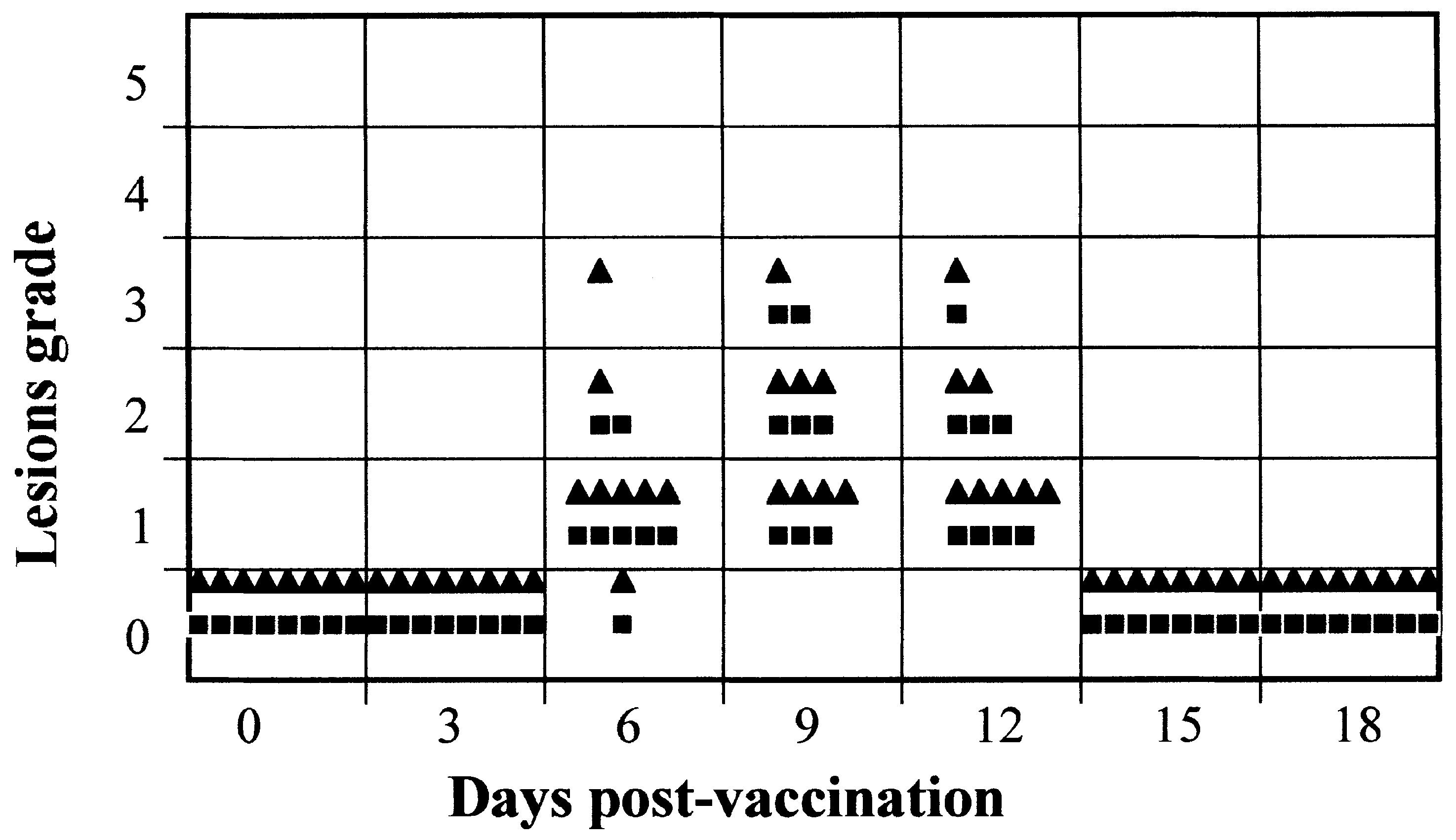
Fig. 3. Eects of administration of 6918VP60-T2 virus after 10 pas-
pressed rabbits. Groups of eight rabbits treated (R) or untreated (Q)
sages in vivo. Groups of eight rabbits were inoculated by s.c. route
with prednisolone were inoculated by i.d. route with 104 pfu of
with Passage 0 (Q) or Passage 10 (R) 6918VP60-T2 virus. Rabbits
6918VP60-T2 virus. Rabbits were observed daily for a period of 18
were observed daily for a period of 18 days and the clinical signs
days and the clinical signs due to virus infection of each animal were
due to virus infection of each animal were ranked from 0 to 6
ranked from 0 to 6 according to Table 1.
3.3. Eects induced by the administration of 6918VP60-
virus-infected does showed any symptomatology as-
To evaluate the eects of recombinant virus infec-
3.4. Analysis of the biological stability of 6918VP60-T2
tion on reproduction, pregnant does were inoculated
at dierent times of gestation (days 7, 14, 21 and 28)
by s.c. route. The daily observation of the animals
The biological stability of the recombinant virus,
showed a total absence of general clinical symptoms in
and therefore its potential to evolve to a virulent state
all inoculated animals. Reproductive parameters such
were evaluated by comparing the eects of rabbit
as number of animals born alive per litter, number of
infection with ``Passage 0'' virus (the same virus stock
animals born dead per litter, number of living animals
used in all the experiments reported in this paper),
per litter 8 dpp, and average weight of each litter at 8
with the eects of rabbit infection with the virus
dpp, for both ®rst and second parturition, have been
obtained after 10 serial passages in rabbits (Passage 10
summarised in Table 4. The overall results showed
virus). Fig. 3 shows a graphic representation of the
that recombinant virus infection did not induce any
symptomatology observed in rabbits infected with
alteration during reproduction. Pregnant does infected
either Passage 0 or Passage 10 virus, according to the
at dierent days of gestation showed reproductive
ranking of myxomatosis clinical signs established in
values being in the expected range for rabbits, and no
Table 1. Rabbits infected with Passage 10 virus exhib-
dierences were observed when recombinant virus-
ited the same mild clinical signs as those infected with
infected does were compared with control does inocu-
Passage 0 virus. Symptoms appeared 5±7 dpi and com-
lated with PBS at the same day of gestation. The
pletely resolved by 15 dpi in both cases. None of the
absence of alterations in reproductive parameters was
infected rabbits exhibited classical severe myxomatosis
maintained in the following parturition (Table 4). Fur-
symptoms. Table 5 shows temperature increases from
thermore, none of the rabbits born from 6918VP60-T2
0 to 2 dpi and from 0 to 4 dpi, as well as weight
Eects induced by 6918VP60-T2 virus infection in immunosuppressed rabbits
Vaccination route Mean body temperature increase
Mean body weight increase Mean antibody titre
J.M. Torres et al. / Vaccine 19 (2001) 174±182
increases from day 0 to 21. No signi®cant dierences
oral vaccination is being used to control enzootic syl-
in body temperature increase or body weight increase
vatic rabies in Europe and North America by means
were observed when rabbits infected with Passage 10
of a recombinant vaccinia-rabies vaccine delivered by
virus were compared with rabbits infected with Passage
baiting [26]. An alternative strategy is the use of
0 virus or control uninfected rabbits. The humoral re-
``transmissible vaccines'', i.e., viral vectors capable of
sponses elicited by rabbits infected with Passage 0 or
spreading within an animal population. Hopefully, the
Passage 10 virus were similar. All infected rabbits
administration of a recombinant vaccine of this
developed high anti-MV and anti-RHDV antibody
characteristics to a small number of captured individ-
uals, would eventually lead to the immunization of a
The genomic stability of 6918VP60-T2 virus was
fraction of animals within a given population, which is
analysed by PCR using oligonucleotide primers exter-
sucient to reduce the spread of the target disease.
nal to the insertion site of the VP60 gene. After 10
This approach might be useful, especially when the dis-
serial passages in rabbits, a product of 3.3 kb (the
tribution, size, and turnover rate of a population pre-
expected size for the recombinant virus) was ampli®ed
cludes capture or baiting techniques as the only means
by PCR with no detection of the corresponding wild-
for antigen delivery. The European rabbit is an
type MV 1.0 kb product (not shown), indicating that
example of such a population. With this in mind, we
the VP60 gene was stably integrated in the MV gen-
have developed a transmissible vaccine against both
myxomatosis and RHD based on a recombinant MV-
VP60 virus capable of spreading through rabbit popu-
lations [25]. The results obtained under laboratory
conditions suggest the recombinant virus might be
eective for wild rabbit immunization. However, since
A number of vaccines are available to protect rab-
the proposed use of 6918VP60-T2 involves the en-
bits against myxomatosis and RHD [4,5,14] which are
vironmental release of a recombinant virus, consider-
useful for immunizing domestic rabbits. However, con-
ations regarding safety issues are as important as the
trol of both diseases among wild rabbit populations
potential ecacy of the candidate vaccine. It is for this
remains an unsolved problem of great concern. In this
reason that safety concerns have been at the core of
regard it should be noted that the European rabbit
the rational design of the proposed immunization
plays a key ecological role in Mediterranean ecosys-
tems. In addition, rabbits are among the most import-
The biological characteristics of MV make it a
ant small game species in several European countries.
good candidate as a vaccine vector in terms of
Immunization of wildlife is dicult to achieve
safety considerations. MV exhibits a very restricted
because direct delivery of vaccines to free ranging ani-
host range, infecting exclusively rabbits (both Sylvi-
mals is not possible. The oral route is considered a
lagus and Oryctolagus spp.). The virus has been
feasible way of vaccine administration. For example,
widely distributed throughout Europe, Australia and
Eects induced by 6918VP60-T2 virus infection in pregnant does
J.M. Torres et al. / Vaccine 19 (2001) 174±182
the Americas for nearly 50 years with no evidence
of infection of other species. Thus, the host
restricted nature of MV minimises the risk of
recombinant vaccine spreading to non-target species
in nature. On the other hand, given the current
widespread geographic distribution of MV, which is
similar to the distribution of RHDV, the ®eld use
of a recombinant MV-VP60 vaccine would normally
not involve the introduction of a virus species that
does not already exist in a particular area.
Safety aspects were also considered in the choice
of the parental MV strain. It was decided not to
use one of the available vaccinal strains, obtained
by cell culture-attenuation of virulent MV strains
[5], as this would involve the release of a new
strain to the environment, which might undergo
reversion to virulence in nature. Instead, we decided
to use an attenuated MV ®eld strain which was
already circulating among wild rabbit populations.
Strain 6918 was selected from a ®eld survey of MV
strains circulating in Spain, which were analysed for
virulence and transmissibility [24]. This strain exhib-
ited adequate biological characteristics for the devel-
opment of a recombinant transmissible vaccine, as
it caused a non-pathogenic infection comparable to
that of cell culture-attenuated vaccinal strains, yet
retaining the capacity of horizontal spreading [24].
Since preservation of the valuable biological prop-
erties of 6918 strain was of major importance in
the development of the recombinant virus, the
foreign gene was inserted in the intergenic site
between ORFs MJ2 and MJ2a, as recombinant
MVs with insertions at this site have been shown
to retain overall parental biological characteristics
[27]. Moreover, the VP60 expression cassette was
inserted into the MV genome using the TDS two-
step selection system [28]. This procedure enables
the construction of recombinant poxviruses without
any marker genes inserted in the ®nal recombinant
viral genome. Thus, the recombinant 6918VP60-T2
does not harbour selectable markers such as anti-
biotic resistance genes, the widespread of which is
currently regarded as a major health and environ-
mental threat. Considering the potential risks associ-
ated with the DNA sequence inserted, it should be
noted that the VP60 gene has been cloned in a
wide range of heterologous systems[15±23] and no
indication of toxicity or side eects associated to
the expression of VP60 have been reported.
Previous results indicated that administration of
either 6918 MV or recombinant 6918VP60-T2 virus to
healthy rabbits under laboratory conditions by stan-
dardised procedures is safe, as all rabbits exhibited
only mild clinical symptoms and rapidly recovered
[24,25]. In this report we have extended the safety
assessment of the vaccine by analysing the potential
J.M. Torres et al. / Vaccine 19 (2001) 174±182
risks of vaccine administration under a varied range of
On the basis of the results previously reported
situations that might occur if the recombinant virus is
[24,25] and those presented in this paper, along with
used for large-scale ®eld immunization of rabbits.
experimental data addressing further safety and e-
Concerning vaccine dosage and the possibility of
cacy issues (to be published elsewhere), the recombi-
accidental administration of an overdose, the results
nant 6918VP60-T2 has been subjected to the
demonstrated vaccine safety even when a 100-fold
mandatory risk assessment process relative to the
overdose (106 PFU) was inoculated (Fig. 1, Table 2).
release of genetically-modi®ed organisms. A limited
Assessment of vaccine eects in immunosuppressed
®eld trial authorised by the Spanish competent auth-
rabbits was considered relevant, given the incidence in
orities is in course. This trial will assess the ecacy
nature of immunocompromised individuals due to
and safety of the vaccine under controlled ®eld con-
infections, environmental or genetic causes. For this
ditions, in the perspective of its use in a large-scale
reason we assayed the eect of vaccine administration
program for the control of myxomatosis and RHD
in rabbits treated with prednisolone, a potent immuno-
suppressor. This treatment induces depletion of circu-
lating eosinophils and mononuclear cells, causing a
strong decrease of the T-cell response with only a
slight eect on B-cell function [29]. It is a commonly
used procedure for the safety evaluation of veterinary
This work was supported by an agreement between
vaccines [30±32]. Results showed that prednisolone
the ``FundacioÂn para el Estudio y Defensa de la Nat-
treated rabbits exhibited similar symptoms to those
uraleza y la Caza'' (FEDENCA) and the ``Instituto
observed in control rabbits (Fig. 2, Table 3). The only
Nacional de InvestigacioÂn y TecnologõÂa Agraria y Ali-
remarkable observation was that immunosuppressed
rabbits showed a subtle tendency to delay the resol-
ution of local lesions: 16±18 dpi vs. 15 dpi (Fig. 2).
Another important aspect addressed was the eect of
6918VP60-T2 virus infection in reproduction. Results
showed that recombinant virus inoculation did not
[1] Murphy, FA, Fauquet, CM, Bishop, DHL, Ghabrial, SA,
alter the reproduction parameters and none of the rab-
Jarvis, AW, Martelli, GP, Mayo, MA, Summers, MD., Virus
bits born from vaccinated does showed myxomatosis-
taxonomy: classi®cation and nomenclature of viruses. Sixth
report of the International Commitee for the Taxonomy of
associated clinical signs (Table 4). In conclusion, the
Viruses. Arch. Virol. 1995;10(Supplement):586 pp. Vienna,
overall results obtained demonstrate a notable lack of
adverse eects attributable to the recombinant virus,
[2] Fenner F, Ross J. Myxomatosis. In: Thompson HV, King CM,
regardless of dose, route or life history stage of indi-
editors. The European rabbit. The history and biology of a suc-
viduals (i.e., neonate, young, pregnant does or immu-
cessful coloniser. Oxford: Oxford University Press, 1994. p.
[3] Kerr Pj, Best SM. Myxoma virus in rabbits. Rev Sci Tech O
Finally the biological stability of the recombinant
virus was analysed. The environmental release of
[4] Fenner F, Woodroofe GM. Protection of laboratory rabbits
recombinant 6918VP60-T2 virus would involve a cer-
against myxomatosis by vaccination with ®broma virus. Aust J
tain number of serial passages in its natural host, even
[5] Saurat P, Gilbert Y, GanieÁre JP. Etude d'une souche de virus
when this capability seemed to be limited to only two
myxomateux modi®e. Rev Med Vet 1978;129:415±51.
serial passages under laboratory conditions [25].
[6] Liu SJ, Xue HP, Pu BQ, Qian SH. A new viral disease in rab-
Should there be a tendency for the virus to evolve to a
bits. Anim Husb Vet Med 1984;16:253±5.
virulent state, serial passage in rabbits would cause it
[7] Morise JP, Le Gall G, Boilleot E. Hepatitis of viral origin in
to do so. Accordingly, the biological stability of
leporidae: introduction and aetiological hypotheses. Rev Sci
6918VP60-T2 was studied by subjecting the virus to 10
[8] Ohlinger VF, Thiel HJ. Rabbit haemorrhagic disease (RHD):
serial passages in rabbits, and the results obtained
characterization of the causative calicivirus. Vet Res
(Fig. 3, Table 5) indicated the recombinant virus main-
tained grossly the same biological characteristics
[9] Villafuerte R, Calvete C, Blanco JC, Lucientes J. Incidence of
through the passages. Thus, the attenuated nature of
viral haemorrhagic disease in wild rabbit populations in Spain.
6918VP60-T2 seems to be a stable trait. On the other
[10] Chasey D. Rabbit haemorrhagic disease: the new scourge of
hand, the genetic analysis indicated that the VP60 gene
Oryctolagus cuniculus. Lab Anim 1997;31:33±44.
remained stably integrated in the MV genome after
[11] Marchandeau S, Chantal J, Portejoie Y, Barraud S, Chaval Y.
serial passage in rabbits, in agreement with the pre-
Impact of viral haemorrhagic disease on a wild population of
viously reported results obtained after 15 serial pas-
European rabbits in France. J Wildl Dis 1998;34:429±35.
[12] Mutze G, Cooke B, Alexander P. The initial impact of rabbit
sages of 6918VP60-T2 virus in RK-13 cell monolayers
haemorrhagic disease on European rabbit populations in South
Australia. J Wildl Dis 1998;34:221±7.
J.M. Torres et al. / Vaccine 19 (2001) 174±182
[13] Pringle CR. Virus taxonomy Ð San Diego. Arch Virol
[23] CastanÄoÂn S, MarõÂn MS, MartõÂn-Alonso JM, Boga JA, Casais
R, Humara JM, OrdaÂs RJ, Parra F. Immunization with potato
[14] ArguÈello JL. Viral haemorrhagic disease of rabbits: vaccination
plants expressing VP60 protein protects against rabbit haemor-
and immune response. Rev Sci Tech O Int Epiz 1991;10:471±
rhagic disease virus. J Virol 1999;73:4452±5.
[24] BaÂrcena J, PageÁs-Mante A, March R, Morales M, RamõÂrez
[15] Boga JA, Casais R, MarõÂn MS, MartõÂn-Alonso JM, CaÂrmenes
MA, SaÂnchez-VizcaõÂno JM, Torres JM. Isolation of an attenu-
RS, Prieto M, Parra F. Molecular cloning, sequencing and ex-
ated myxoma virus ®eld strain that confers horizontal transmis-
pression in Escherichia coli of the capsid protein gene from rab-
sible protection against myxomatosis on contacts of vaccinates.
bit haemorrhagic disease virus (Spanish isolate AST/89). J Gen
[25] BaÂrcena J, Morales M, VaÂzquez B, Boga JA, Parra F, Lucientes
[16] Laurent S, Vautherot JF, Madelaine MF, Le Gall G,
J, PageÁs-Mante A, SaÂnchez-VizcaõÂno JM, Blasco R, Torres JM.
Rasschaert D. Recombinant rabbit haemorrhagic disease virus
Horizontal transmissible protection against myxomatosis and
capsid protein expressed in baculovirus self-assembles into virus-
rabbit haemorrhagic disease using a recombinant myxoma
like particles and induces protection. J Virol 1994;68:6794±8.
[17] Plana-Duran J, Bastons M, Rodriguez MJ, Climent I, CorteÂs E,
[26] Brochier B, Aubert MF, Pastoret PP, Masson E, Schon J,
Vela C, Casal I. Oral immunization of rabbits with VP60 par-
Lombard M, Chappuis G, Languet B, Desmettre P. Field use
ticles confers protection against rabbit haemorrhagic disease.
of a vaccinia-rabies recombinant vaccine for the control of syl-
vatic rabies in Europe and North America. Rev Sci Tech O
[18] Sibilia M, Boniotti MB, Angoscini P, Capucci L, Rossi C. Two
independent pathways of expression lead to self-assembly of the
[27] Jackson RJ, Hall DF, Kerr PJ. Construction of recombinant
rabbit haemorrhagic disease virus capsid protein. J Virol
myxoma viruses expressing foreign genes from dierent inter-
genic sites without associated attenuation. J Gen Virol
[19] Bertagnoli S, Gel® J, Petit F, Vautherot JF, Rasschaert D,
Laurent S, Gall G, Boilletot E, Chantal J, Boucraut-Baralon C.
[28] Falkner FG, Moss B. Transient dominant selection of recombi-
Protection of rabbits against rabbit viral haemorrhagic disease
nant vaccinia viruses. JVirol 1990;64:3108±11.
with a vaccinia-RHDV recombinant virus. Vaccine 1996;14:506±
Immunosuppressive eect of corticosteroids on rabbit's humoral
[20] Bertagnoli S, Gel® J, Gall G, Boilletot E, Vautherot JF,
and cellular response. Allergol Immunopathol (Madr)
Rasschaert D, Laurent S, Petit F, Boucraut-Baralon C, Milon
A. Protection against myxomatosis and rabbit viral haemorrha-
[30] ArguÈello JL. ContribucioÂn a la pro®laxis de la mixomatosis del
gic disease with recombinant myxoma viruses expressing rabbit
conejo mediante el uso de una cepa homoÂloga. Medicina
[31] Ciuchini F, Pestalozza S, Buonavoglia C, Di Trani L, Tollis
[21] Fischer L, Le Gros FX, Mason PW, Paoletti E. A recombinant
M, Orfei Z. Eects of corticosteroids mediated immunosup-
canarypox virus protects rabbits against a lethal rabbit haemor-
pression on the distribution of rabies vaccine virus in red
rhagic disease virus (RHDV) challenge. Vaccine 1997;15:90±6.
foxes orally immunized against rabies. J Vet Med B
[22] Boga JA, MartõÂn-Alonso JM, Casais R, Parra F. A single dose
immunization with rabbit haemorrhagic disease virus major cap-
[32] Pedersen NC. Immunogenicity and ecacy of a commercial
sid protein produced in Saccharomyces cerevisiae induces pro-
feline leukemia virus vaccine. J Vet Intern Med 1993;7:
tection. J Gen Virol 1997;78:2315±8.
This chemical compatibility guide was assembled from known compatibility data for PTFE materials andshould be used only as a general guide for determining the suitability ofGORE sealants for specific applications. An independent study of the compatibility with your specificfluids is advised for confirmation of chemical compatibility. When immersion tests are performed withGORE sealants, the test s
UTAH DEPARTMENT OF HEALTH (UDOH) GUIDANCE FOR THE MANAGEMENT OF PERSONS ON TREATMENT FOR LATENT TUBERCULOSIS (TB) INFECTION (LTBI) WITH ISONIAZID (INH) AND RIFAPENTINE (RPT) BY DOT, WEEKLY FOR On December 9, 2011, The Centers for Disease Control and Prevention published the following article in the Morbidity and Mortality Weekly Report, Vol. 60, No. 48, pp. 1650-1653: Recommenda
 Safety evaluation of a recombinant myxoma-RHDV virus
inducing horizontal transmissible protection against myxomatosis
Juan M. Torresa,*, Miguel A. RamõÂreza, MoÂnica Moralesa, Juan BaÂrcenaa,
BeleÂn VaÂzqueza, Enric EspunÄab, Albert PageÁs-ManteÂb, Jose M. SaÂnchez-VizcaõÂnoa
aCentro de InvestigacioÂn en Sanidad Animal (CISA-INIA), Valdeolmos, 28130 Madrid, Spain
Received 14 February 2000; received in revised form 9 May 2000; accepted 12 May 2000
We have recently developed a transmissible vaccine to immunize rabbits against myxomatosis and rabbit haemorrhagic disease
based on a recombinant myxoma virus (MV) expressing the rabbit haemorrhagic disease virus (RHDV) capsid protein [BaÂrcena
et al. Horizontal transmissible protection against myxomatosis and rabbit haemorragic disease using a recombinant myxoma
virus. J. Virol. 2000;74:1114±23]. Administration of the recombinant virus protects rabbits against lethal RHDV and MV
challenges. Furthermore, the recombinant virus is capable of horizontal spreading promoting protection of contact animals, thus
providing the opportunity to immunize wild rabbit populations. However, potential risks must be extensively evaluated before
considering its ®eld use. In this study several safety issues concerning the proposed vaccine have been evaluated under
laboratory conditions. Results indicated that vaccine administration is safe even at a 100-fold overdose. No undesirable eects
were detected upon administration to immunosuppressed or pregnant rabbits. The recombinant virus maintained its attenuated
phenotype after 10 passages in vivo. 7 2000 Elsevier Science Ltd. All rights reserved.
Safety evaluation of a recombinant myxoma-RHDV virus
inducing horizontal transmissible protection against myxomatosis
Juan M. Torresa,*, Miguel A. RamõÂreza, MoÂnica Moralesa, Juan BaÂrcenaa,
BeleÂn VaÂzqueza, Enric EspunÄab, Albert PageÁs-ManteÂb, Jose M. SaÂnchez-VizcaõÂnoa
aCentro de InvestigacioÂn en Sanidad Animal (CISA-INIA), Valdeolmos, 28130 Madrid, Spain
Received 14 February 2000; received in revised form 9 May 2000; accepted 12 May 2000
We have recently developed a transmissible vaccine to immunize rabbits against myxomatosis and rabbit haemorrhagic disease
based on a recombinant myxoma virus (MV) expressing the rabbit haemorrhagic disease virus (RHDV) capsid protein [BaÂrcena
et al. Horizontal transmissible protection against myxomatosis and rabbit haemorragic disease using a recombinant myxoma
virus. J. Virol. 2000;74:1114±23]. Administration of the recombinant virus protects rabbits against lethal RHDV and MV
challenges. Furthermore, the recombinant virus is capable of horizontal spreading promoting protection of contact animals, thus
providing the opportunity to immunize wild rabbit populations. However, potential risks must be extensively evaluated before
considering its ®eld use. In this study several safety issues concerning the proposed vaccine have been evaluated under
laboratory conditions. Results indicated that vaccine administration is safe even at a 100-fold overdose. No undesirable eects
were detected upon administration to immunosuppressed or pregnant rabbits. The recombinant virus maintained its attenuated
phenotype after 10 passages in vivo. 7 2000 Elsevier Science Ltd. All rights reserved.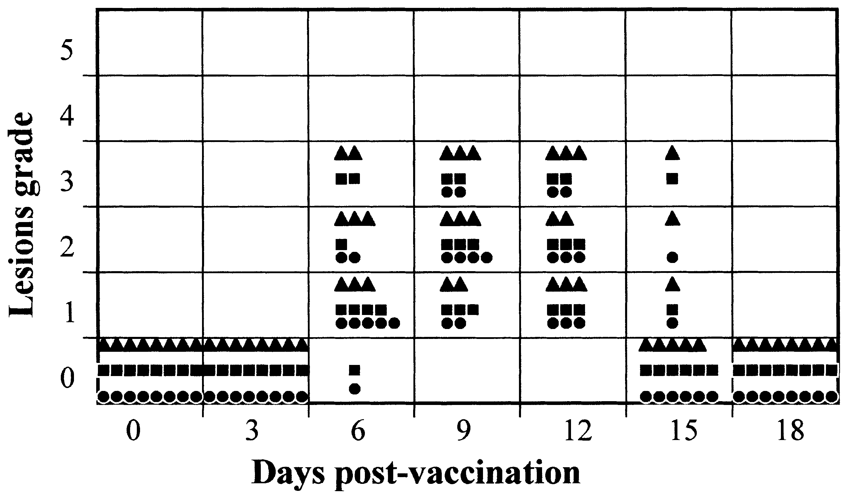 J.M. Torres et al. / Vaccine 19 (2001) 174±182
virus. Control rabbits were vaccinated but not treated
with prednisolone. Rabbits were observed daily for a
period of 21 days and clinical symptoms were
Data were analysed using a Student's t-test for non-
recorded. Weight and temperature determinations were
paired variants. Signi®cance was considered when p `
made on each animal until the 21st day. Serum
samples extracted 0 and 21 days after immunization
were used to evaluate the serological responses against
MV and RHDV by ELISA. Antibody titres were
3.1. Eects induced by the administration of an overdose
2.5. Administration of 6918VP60-T2 virus to pregnant
Previous work showed 104 pfu was an ecient vac-
cine dose to ensure horizontal transmissible protection
Groups of six pregnant does were inoculated at
against myxomatosis and RHD, either by direct con-
dierent times of gestation (days 7, 14, 21 and 28) by
tact or in a ¯ea-mediated process [25]. To evaluate the
s.c. route at the back with 104 pfu of 6918VP60-T2
eects of administering an overdose of the vaccine,
virus. Control does were inoculated at the same days
wild rabbits were inoculated by i.d. or s.c. route with
of gestation with 0.5 ml of phosphate-buered saline
dierent doses of 6918VP60-T2 virus (104, 105 and 106
(PBS). Animals were observed daily and general clini-
cal symptoms were recorded. No body weight and
In order to obtain a semi-quantitative measure to
temperature determinations were performed in order
allow graphic representation and objective comparison,
to minimise the handling-induced stress in does, which
the classical myxomatosis symptoms were classi®ed in
are specially sensible during gestation. The following
a ranking of 1 to 6 points (see Table 1), and the results
reproductive parameters were recorded both at ®rst
registered during the observation period were rep-
and second parturition: number of animals born alive
resented (Fig. 1). Rabbits inoculated by i.d. route dis-
per litter; number of animals born dead per litter;
played similar clinical signs at all vaccine doses tested.
J.M. Torres et al. / Vaccine 19 (2001) 174±182
virus. Control rabbits were vaccinated but not treated
with prednisolone. Rabbits were observed daily for a
period of 21 days and clinical symptoms were
Data were analysed using a Student's t-test for non-
recorded. Weight and temperature determinations were
paired variants. Signi®cance was considered when p `
made on each animal until the 21st day. Serum
samples extracted 0 and 21 days after immunization
were used to evaluate the serological responses against
MV and RHDV by ELISA. Antibody titres were
3.1. Eects induced by the administration of an overdose
2.5. Administration of 6918VP60-T2 virus to pregnant
Previous work showed 104 pfu was an ecient vac-
cine dose to ensure horizontal transmissible protection
Groups of six pregnant does were inoculated at
against myxomatosis and RHD, either by direct con-
dierent times of gestation (days 7, 14, 21 and 28) by
tact or in a ¯ea-mediated process [25]. To evaluate the
s.c. route at the back with 104 pfu of 6918VP60-T2
eects of administering an overdose of the vaccine,
virus. Control does were inoculated at the same days
wild rabbits were inoculated by i.d. or s.c. route with
of gestation with 0.5 ml of phosphate-buered saline
dierent doses of 6918VP60-T2 virus (104, 105 and 106
(PBS). Animals were observed daily and general clini-
cal symptoms were recorded. No body weight and
In order to obtain a semi-quantitative measure to
temperature determinations were performed in order
allow graphic representation and objective comparison,
to minimise the handling-induced stress in does, which
the classical myxomatosis symptoms were classi®ed in
are specially sensible during gestation. The following
a ranking of 1 to 6 points (see Table 1), and the results
reproductive parameters were recorded both at ®rst
registered during the observation period were rep-
and second parturition: number of animals born alive
resented (Fig. 1). Rabbits inoculated by i.d. route dis-
per litter; number of animals born dead per litter;
played similar clinical signs at all vaccine doses tested.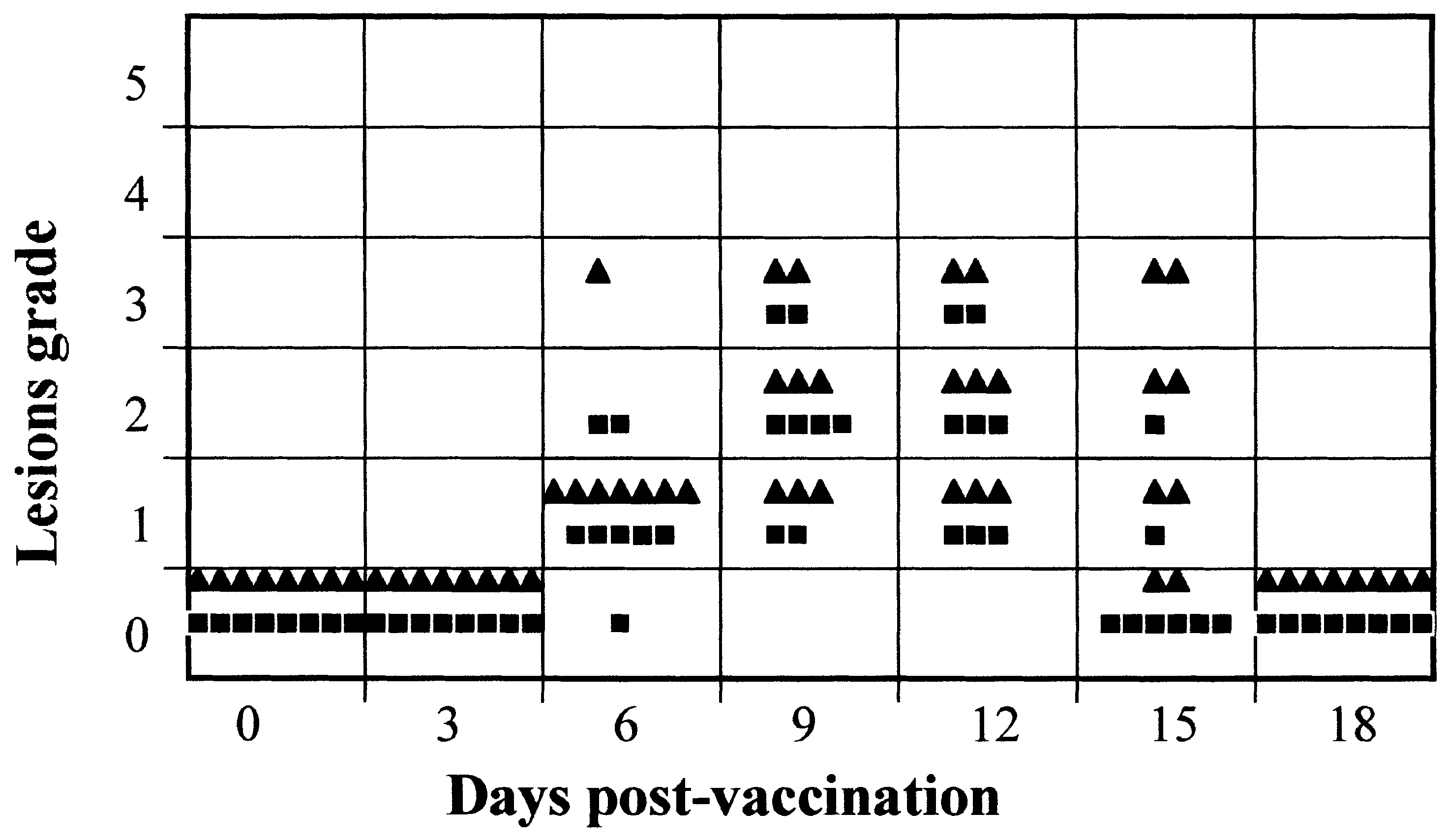
 J.M. Torres et al. / Vaccine 19 (2001) 174±182
Fig. 2. Eects of administering 6918VP60-T2 virus to immunosup-
Fig. 3. Eects of administration of 6918VP60-T2 virus after 10 pas-
pressed rabbits. Groups of eight rabbits treated (R) or untreated (Q)
sages in vivo. Groups of eight rabbits were inoculated by s.c. route
with prednisolone were inoculated by i.d. route with 104 pfu of
with Passage 0 (Q) or Passage 10 (R) 6918VP60-T2 virus. Rabbits
6918VP60-T2 virus. Rabbits were observed daily for a period of 18
were observed daily for a period of 18 days and the clinical signs
days and the clinical signs due to virus infection of each animal were
due to virus infection of each animal were ranked from 0 to 6
ranked from 0 to 6 according to Table 1.
J.M. Torres et al. / Vaccine 19 (2001) 174±182
Fig. 2. Eects of administering 6918VP60-T2 virus to immunosup-
Fig. 3. Eects of administration of 6918VP60-T2 virus after 10 pas-
pressed rabbits. Groups of eight rabbits treated (R) or untreated (Q)
sages in vivo. Groups of eight rabbits were inoculated by s.c. route
with prednisolone were inoculated by i.d. route with 104 pfu of
with Passage 0 (Q) or Passage 10 (R) 6918VP60-T2 virus. Rabbits
6918VP60-T2 virus. Rabbits were observed daily for a period of 18
were observed daily for a period of 18 days and the clinical signs
days and the clinical signs due to virus infection of each animal were
due to virus infection of each animal were ranked from 0 to 6
ranked from 0 to 6 according to Table 1.