La tétracycline, connue sous le nom commercial Sumycin, agit en bloquant la fixation de l’ARNt sur la sous-unité 30S ribosomale, interrompant l’élongation de la chaîne protéique bactérienne. Ce mécanisme confère une activité sur un spectre large, incluant bactéries Gram positives, Gram négatives, rickettsies et spirochètes. Sa biodisponibilité digestive varie selon la prise alimentaire et les interactions avec les ions divalents comme calcium et magnésium. Sa diffusion tissulaire est importante, notamment dans les voies respiratoires et génito-urinaires. L’élimination se fait par voie rénale et biliaire. Les effets indésirables incluent photosensibilisation, troubles digestifs et coloration dentaire en cas d’administration précoce. Les guides thérapeutiques mentionnent sumycin prix, en soulignant la nécessité de restreindre son utilisation afin de limiter les résistances acquises.
Doi:10.1016/j.exer.2005.11.012
Experimental Eye Research 83 (2006) 128e132
Effect of viagra on retinal vein diameter in AMD patients
Tatyana I. Metelitsina, Juan E. Joan C. DuPont,
Scheie Eye Institute, Department of Ophthalmology, University of Pennsylvania, 51 North 39th Street, Philadelphia, PA 19104, USA
Received 28 July 2005; accepted in revised form 4 November 2005
The aim of the present study was to investigate the effect of sildenafil citrate (viagra) on retinal venous diameter in patients with age-relatedmacular degeneration (AMD). We investigated 14 male patients in a double-masked, randomized, placebo-controlled, crossover study. In eachsubject, one eye with typical non-exudative AMD fundus features was studied. Each of the subjects received 100 mg dose of sildenafil or match-ing placebo on two separate study visits. Monochromatic fundus photographs were obtained in the study eye before dosing and then 30, 90, 180and 300 min later. Measurements of the diameter of the major retinal veins from digitized negatives were carried out using ‘‘Vessel map’’ staticvessel analysis program (IMEDOS GmbH, Weimar, Germany). Statistical analysis of the data comparing the effect of sildenafil and placebo onvenous diameters was performed using analysis of variance (ANOVA) for repeated measures. An analysis of variance (ANOVA) comparing theeffects of sildenafil citrate and placebo on retinal vein diameters showed a significant interaction between time and treatment (P ¼ 0.03). Incomparison to placebo, sildenafil citrate produced a statistically significant vasodilatation of major retinal veins of 4.7% at 90 min(P ¼ 0.004), 5.5% at 180 min (P < 0.0001) and 5.8% at 300 min (P < 0.0001). At 30 min there was a 2.2% difference, which was not statis-tically significant (P ¼ 0.14). Our results suggest that in patients with age-related macular degeneration, sildenafil citrate (viagra) produces a sta-tistically significant vasodilatation of major retinal veins that is similar to what has been reported in normal subjects. Whether this vasodilatationis associated with changes in retinal blood flow needs to be further investigated. Ó 2006 Elsevier Ltd. All rights reserved.
Keywords: sildenafil citrate (viagra); retinal vein diameter; age-related macular degeneration (AMD)
oxide (NO) (produce smooth muscle relaxation and dilatation of
Sildenafil citrate (viagra), the first oral drug approved for
blood vessels due to the increase in levels of cGMP (
the treatment of erectile dysfunction, selectively inhibits phos-
phodiesterase 5 (PDE5), the isozyme that metabolizes cyclic
). By blocking PDE5 sildenafil increases the levels
guanosine monophosphate (c-GMP) in the corpus cavernosum
of c-GMP and thus greatly enhances the dilating effects of NO
smooth muscle, PDE5 is also found in other human tissues
This vasodilatory quality of sildenafil is of great interest
because of the potential use of this type of compound in the
). Endothelium-derived relaxing factors, such as nitric
treatment of vascular occlusive disease. Indeed several inves-tigators have studied the effects of sildenafil on the retinalvessel diameter in normal subjects (
* Corresponding author. Tel.: þ1 215 662 8039; fax: þ1 215 662 8025.
by sildenafil in the human retina was reported by some studies
(J.C. DuPont), (G.-shuang Ying), (C. Liu).
0014-4835/$ - see front matter Ó 2006 Elsevier Ltd. All rights reserved. doi:10.1016/j.exer.2005.11.012
T.I. Metelitsina et al. / Experimental Eye Research 83 (2006) 128e132
In this study we investigate the effect of sildenafil on the
of either 100 mg of sildenafil citrate (Viagra; Pfizer Inc,
retinal vessel diameter in patients with age-related macular de-
New York) or matching placebo on the first study visit. The
alternative drug was tested on the second study visit. Placebo
pills were identical to the sildenafil ones, but they did not con-
have suggested that impairment of the choroidal circu-
tain the active component. The same protocol was performed
lation may play an important role in the etiology of this dis-
on both study days, which were separated by a wash out period
ease. Little is known however, about the retinal vasculature
of three or more days. In order to prevent bias, both patients
changes in this disease. has shown using la-
and investigators were masked to the treatment modalities.
ser Doppler velocimetry that pulsatily in retinal arteries is
After pupillary dilation was achieved with tropicamide 1%
higher in patients with AMD than healthy controls. The exact
(Alcon, Fort Worth, TX) and phenylephrine hydrochloride
implications of this finding are not known but suggest that per-
10% (Sanofi Winthrop, New York, NY), monochromatic fun-
fusion abnormalities go beyond the choroid in patients with
dus photographs (l ¼ 570 nm) were obtained with a Zeiss fun-
age-related macular degeneration. Because the vasculature of
dus camera (Oberkochen, Germany) on Kodak Plus-X pan
the fundus seems to be affected by AMD it is important to
film (Rochester, New York, USA). Photos of the study eye
study whether sildenafil affects the vasculature of AMD pa-
of each patient were obtained while the subjects were seated
in the darkened room. Intraocular pressure (IOP), brachial ar-
show any statistically significant effect of sildenafil on the
tery systolic and diastolic blood pressure (BPs and BPd) and
choroid of AMD patients. In this study we investigate the ef-
heart rate (HR) were obtained immediately after the photo-
fect of sildenafil on the retinal vasculature of AMD patients.
graphs were taken. Tono-pen XL (Mentor Ophthalmics, Nor-well, MA) and automated sphygmomanometer (Accutorr 1A,
Datascope, Paramus, NJ) were used to measure IOP and BP,respectively. The mean brachial artery pressure (BPm) was
Fourteen male AMD patients (13 Caucasians and 1 Afri-
calculated according to the following formula:
can-American) with a mean age of 75 Æ 7 years (Æ1SD)were included in this double-masked, randomized, placebo-
controlled, crossover study. AMD features of enrolled patients
Perfusion pressure (PP) for the study eye was estimated
were similar or worse than those present in eyes graded as
AMD category 3 in the AREDS study. Only one eye of eachpatient was included in the study. Study eyes had clear ocular
media, intraocular pressure (IOP) of 21 mm Hg or less, pupil-lary dilatation of 5 mm or more, steady fixation, no intraocular
All tests mentioned above were performed prior to the
diseases other than AMD and no choroidal neovascularization
administration of the drug, and then 30, 90, 180 and 300 min,
(CNV). Right eyes were chosen in ten patients and left eyes in
thereafter. These times were chosen to coincide with the
the other four. All study eyes had large drusen and eight eyes
maximal serum concentration levels of sildenafil, which are
had retinal pigment epithelium hyperpigmentary changes. Five
reached in 30e60 min. Plasma half-life of sildenafil is about
patients had small areas of extrafoveal geographic atrophy in
the study eye or the fellow eye. Two patients had exudative
Approximately ten fundus photographs were obtained at
AMD with a disciform scar present in the fellow eye. External
each time point for each patient. Photographic negatives
and slit lamp examinations were normal except for the pres-
were scanned and digitized, using a Nikon SF-2000 35 mm
ence of mild lens nuclear sclerosis in nine study eyes and in-
film scanner (Tokyo, Japan). Out of the ten digital images,
traocular lens implants in three eyes.
five photographs with the sharpest focus were selected for
A history of well-controlled systemic hypertension was re-
further analysis in a masked fashion.
ported in nine patients. All of them were taking antihyperten-
Retinal vein diameter was measured with ‘‘Vessel map’’
sive medications. None of the 14 subjects enrolled in the study
static vessel analysis program software (IMEDOS GmbH,
had a history of systemic hypotension or serious heart condi-
Weimar, Germany). This program enables the determination
tion or was receiving nitrate therapy. All study subjects took
of retinal vessel diameter from digitized photographs. The ves-
the same medications throughout the length of the study, and
sel diameter is determined segment by segment along the
none of the study participants was under fasting conditions.
length of a blood vessel in a predefined measurement window.
The study was carried out after the approval from University
The instrument creates an intensity or brightness profile per-
of Pennsylvania Institutional Review Board. The tenets of Dec-
pendicular to the vessel. The width of the vessel is defined
laration of Helsinki were followed. All subjects signed an appro-
as the distance from wall to wall through the midpoint of
priate IRB approved consent form after the detailed explanations
of the study procedures. We have previously reported our find-
Two reference points were identified on vessel bifurcation
ings on the effect of sildenafil citrate on the choroidal blood
for each photograph (see This allowed us to overlap
all images of one subject assuring that all fundus details
The study design included two separate study visits, with
from one picture corresponded to the same fundus details in
all patients being randomized to receive a single oral dose
all images of the same patient. This preliminary step enabled
T.I. Metelitsina et al. / Experimental Eye Research 83 (2006) 128e132
correlation from multiple veins from the same eye. SAS 9.1software (Cary, North Carolina, USA) was used for the analy-sis of the data.
Mean retinal venous diameters at baseline, 30, 90, 180 and
300 min for sildenafil and placebo treatments are shown in(An analysis of variance (ANOVA) comparingthe effects of sildenafil citrate and placebo on retinal veins di-ameters showed a significant interaction between time andtreatment (P ¼ 0.03), therefore, comparisons of both treat-ments at each time point were performed.
In comparison to placebo, sildenafil citrate caused a statisti-
cally significant vasodilatation of major retinal veins at 90 min(P ¼ 0.004), 180 min (P < 0.0001) and 300 min (P < 0.0001). Compared with the mean of placebo treatment, the mean ve-
Fig. 1. Monochromatic fundus photograph of a typical study eye of an AMD
nous diameter after sildenafil citrate treatment was 4.7% larger
patient with multiple large drusen. Measurement of the inferior temporal veinsegment was performed using the ‘‘Vessel map’’ software. Rectangle shows
at 90 min, 5.5% larger at 180 min, and 5.8% larger at 300 min.
segment of the vessel chosen for measurement. Circles show two reference
There was no statistically significant difference between
points that are used by the software to overlap multiple images.
placebo and sildenafil at 30 min (P ¼ 0.14).
multiple measurements of the same vessel simultaneously on
demonstrated a statistically significant decrease in BPm of
15.2% (P ¼ 0.006) and PP of 22.3% (P ¼ 0.006) at 30 min
A predefined measurement window of about ÿ1/2 disk di-
after administration of sildenafil citrate, we investigated
ameter ensured that the length of the vessel segment measured
whether changes in BPm and PP are associated with an in-
was the same for all images of the same subject (The
crease in venous diameter after sildenafil treatment. Our anal-
same segment of the vessel chosen for analysis (was
ysis, however, showed no statistically significant association
evaluated in all photos of the same patient. To make sure
between changes in retinal vein diameters and changes in
that the location of the measurement window was consistent
BPm (P ¼ 0.07) or PP (P ¼ 0.25). Also no statistically sig-
on all images, a transparent plastic template with an outline
nificant association between changes in retinal vein diameter
of the disk and the major vessels was superimposed on all
and changes in IOP (P ¼ 0.67) after sildenafil were detected
Measurements were performed on straight segments of the
vessels within 1.5 disk diameters from the edge of the optic
nerve head. We avoided vascular bifurcations and arteriove-nous crossings. Major veins were chosen for analysis. In ten
In this current study we investigated the effect of sildenafil
patients, two major veins were measured. In four patients
on retinal vessel diameter in patients with AMD. Sildenafil
only one major vein was analyzed because a second vein in
citrate produced statistically significant dilatation of major
proper focus was not available in all photographs. Thus a total
retinal veins at 90, 180 and 300 min in this group of patients.
number of 24 veins were included in our analysis. All circula-
The effect of sildenafil on the retinal vessel diameters of
tory measurements are shown in arbitrary units (AU).
AMD patients has not been studied before. Our previous study
Statistical analysis of the data comparing the differences in
on the effects of sildenafil on the choroidal circulation of
retinal vessel diameters before and after administration of sil-
AMD patients did not show any statistically significant
denafil and placebo was performed using analysis of variance(ANOVA) for repeated measures. To compare the differences
between these two groups at each time point (baseline, 30,
Venous diameters after administration of sildenafil citrate and placebo at all
90, 180 and 300 min), alpha level adjustments for multiple
comparisons by means of Bonferroni approach were also car-
ried out. Because we had five time points comparisons, we
considered P ¼ 0.01 (0.05/5 ¼ 0.01) as statistically signifi-
cant. Regression analyses between vein diameter changes
from baseline and pressure changes (IOP, PP and BP) from
baseline were also performed and a P value of 0.05 was con-
sidered to be statistically significant. Since the diameters from
two veins were measured for most of the patients, the general-
ized estimating equations (GEE) were used to adjust the
a Correlation from multiple veins of the same eye was adjusted.
T.I. Metelitsina et al. / Experimental Eye Research 83 (2006) 128e132
significant venous dilatation starting at 90 min and the veinsremained dilatated even at 300 min.
group also showed a 5.8% increase in venous and arterialdiameter after administration of 50 mg of sildenafil. Thepeak of the changes, however, was observed at 30 minand by 120 min both arterial and venous diameters had re-turned to baseline values. In contrast to this study, did not observe any statistically significant effecton the retinal arterial diameters, and no effect of sildenafilwas found on a flicker-induced vasodilatation in retinal ar-teries or veins.
Our previous study in normal male volunteers (
did not reveal any statistically significant effectin retinal venous and arterial diameters after administrationof sildenafil. This study, however, used an older method that
Fig. 2. Average retinal venous diameters in mm at baseline, 30 min, 90 min,180 and 300 min after treatment with placebo and sildenafil citrate (viagra).
had only an 80% power to detect 6.5% difference between
Error bars correspond to Æ1 standard error (SE).
the two groups. The lower sensitivity of this older methodmay explain the discrepancy with the other above-mentionedstudies.
differences in the choroidal blood flow parameters after ad-
In conclusion, our results show a statistically significant va-
ministration of sildenafil or placebo at any of the time points
sodilatation of major retinal veins at 90, 180 and 300 min after
administration of a 100 mg of sildenafil citrate in AMD pa-
obtained in these two studies may be due to the fact that the
tients. This effect is similar to what has been previously re-
retinal and choroidal circulations are two different vascular
ported in normal subjects. Whether increased retinal venous
beds that may have different concentration of target receptors.
diameters are associated with increased retinal blood flow in
The significance of this retinal vasodilatation following silde-
AMD patients and whether this effect may be of any therapeu-
nafil treatment in AMD and normal patients is not known at
tic value are questions that need further investigation.
this time. This issue should be further clarified in the futurewhen the effect of sildenafil on retinal blood flow iselucidated.
We also attempted to measure the effect of sildenafil citrate
on retinal arteries. We were able to measure only 11 arteries of11 patients. One of the major problems was that the light reflex
Boolell, M., Allen, M.J., Ballard, S.A., Gepi-Attee, S., Muirhead, G.J.,
present in the center of the arterioles introduced errors in the
Naylor, A.M., Osterloh, I.H., Gingell, C., 1996. Sildenafil: an orally activetype 5 cyclic GMP-specific phosphodiesterase inhibitor for the treatment
diameter measurements and this prevented us from getting
of penile erectile dysfunction. Int. J. Impot. Res. 8, 47e52.
an accurate estimation of arterial diameters in many instances.
Furchgott, R.F., Vanhoutte, P.M., 1989. Endothelium-derived relaxing and
All previous studies of the effect of sildenafil on the retinal
contracting factors. FASEB J. 3, 2007e2018.
vasculature were performed in normal volunteers.
Friedman, E., Ivry, M., Ebert, E., Glynn, R., Gragoudas, E., Seddon, J., 1989.
showed a significant 4.7% increase in the retinal ve-
Increased scleral rigidity and age-related macular degeneration. Ophthal-mology 196, 104e108.
nous diameter at 80 min after 100 mg of sildenafil citrate.
Friedman, E., Krupsky, S., Lane, A.M., Oak, S.S., Friedman, E.S., Egan, K.,
The findings from this study and the timing of the changes ob-
Gragoudas, E.S., 1995. Ocular blood flow velocity in age-related macular
served are very similar to our current data. Maximum serum
degeneration. Ophthalmology 102, 640e646.
Gruetter, C.A., Gruetter, D.Y., Lyon, J.E., Kadowitz, P.J., Ignarro, L.J., 1981.
and it is interesting that we found statistically
Relationship between cyclic guanosine 30, 50-mono-phosphate formationand relaxation of coronary arterial smooth muscle by glyceryl trinitrate,nitroprusside, nitrite and nitric oxide: effects of methylene blue andmethemoglobin. J. Pharmacol. Exp. Ther. 219, 181e186.
Grunwald, J.E., Hariprasad, S.M., DuPont, J., Maguire, M.G., Fine, S.L.,
Association between change in retinal venous diameter and change in mean
Brucker, A.J., Maguire, A.M., Ho, A.C., 1998. Foveolar choroidal blood
intraocular pressure (IOP), blood pressure (BPm), and perfusion pressure (PP)
flow in age-related macular degeneration. Invest. Ophthalmol. Vis. Sci. 39, 385e390.
Grunwald, J.E., Metelitsina, T., Grunwald, L., 2002. Effect of sildenafil citrate
(viagra) on retinal blood vessel diameter. Am. J. Ophthalmol. 133, 809e812.
Grunwald, J.E., Metelitsina, T.I., Dupont, J.C., Ying, G.S., Maguire, M.G.,
2005. Reduced foveolar choroidal blood flow in eyes with increasing
a Estimated from the model: diameter change ¼ intercept þ b  pressure
AMD severity. Invest. Ophthalmol. Vis. Sci. 46, 1033e1038.
Ignarro, L.J., Kadowitz, P.J., 1985. The pharmacological and physiological
b Correlations between repeated measurements from same subjects are
role of cyclic GMP in vascular smooth muscle relaxation. Annu. Rev. Phar-
T.I. Metelitsina et al. / Experimental Eye Research 83 (2006) 128e132
Ignarro, L.J., Buga, G.M., Wood, K.S., Byrns, R.E., Chaudhuri, G., 1987.
Pauleikhoff, D., Chen, J.C., Chisholm, I.H., et al., 1990. Choroidal perfusion
Endothelium-derived relaxing factor produced and released from artery
abnormality with age-related Bruch’s membrane change. Am. J. Ophthal-
and vein is nitric oxide. Proc. Natl. Acad. Sci. U.S.A. 84, 9265e9269.
Marmor, M.F., Kessler, R., 1999. Sildenafil (viagra) and ophthalmology. Surv.
Pfizer. Investigative Brochure. Version 69-5485-00-9.
Polak, K., Wimpissinger, B., Berisha, F., Georgopoulos, M., Schmetterer, L.,
Metelitsina, T.I., Grunwald, J.E., DuPont, J.C., Ying, G., 2005. Effect of viagra
2003. Effects of sildenafil on retinal blood flow and flicker-induced retinal
on the foveolar choroidal circulation of AMD patients. Exp. Eye Res. 81,
vasodilatation in healthy subjects. Invest. Ophthalmol. Vis. Sci. 44,
Pache, M., Meyer, P., Prunte, C., Orgul, S., Nuttli, I., Flammer, J., 2002. Sil-
Sato, E., Feke, G.T., Menke, M.N., Wallace McMeel, J., 2005 Jun 3. Retinal
denafil induces retinal vasodilatation in healthy subjects. Br. J. Ophthal-
haemodynamics in patients with age-related macular degeneration. Eye.
Arbonne Clear Advantage vs Proactiv for Acne - A Review of Two Different Approaches to Acne Treatment Facial skin problems are a major concern for many people. The most common skin problem is acne. It is important to understand why acne happens in the first place in order to effectively combat it. Acne begins at puberty. It is caused by an increase in androgens, which is a male sex hormone.
Y a-t-il des risques liés à l’injection ?Les produits de contraste actuels sont généralement très bien tolérés, cependantl’injection peut entraîner une réaction d’intolérance. Ces réactions imprévisibles Vous allez sont plus fréquentes chez les patients ayant eu une injection mal tolérée d’un deces produits ou ayant des antécédents allergiques. Elles sont généra


 Experimental Eye Research 83 (2006) 128e132
Effect of viagra on retinal vein diameter in AMD patients
Tatyana I. Metelitsina, Juan E. Joan C. DuPont,
Scheie Eye Institute, Department of Ophthalmology, University of Pennsylvania, 51 North 39th Street, Philadelphia, PA 19104, USA
Received 28 July 2005; accepted in revised form 4 November 2005
The aim of the present study was to investigate the effect of sildenafil citrate (viagra) on retinal venous diameter in patients with age-relatedmacular degeneration (AMD). We investigated 14 male patients in a double-masked, randomized, placebo-controlled, crossover study. In eachsubject, one eye with typical non-exudative AMD fundus features was studied. Each of the subjects received 100 mg dose of sildenafil or match-ing placebo on two separate study visits. Monochromatic fundus photographs were obtained in the study eye before dosing and then 30, 90, 180and 300 min later. Measurements of the diameter of the major retinal veins from digitized negatives were carried out using ‘‘Vessel map’’ staticvessel analysis program (IMEDOS GmbH, Weimar, Germany). Statistical analysis of the data comparing the effect of sildenafil and placebo onvenous diameters was performed using analysis of variance (ANOVA) for repeated measures. An analysis of variance (ANOVA) comparing theeffects of sildenafil citrate and placebo on retinal vein diameters showed a significant interaction between time and treatment (P ¼ 0.03). Incomparison to placebo, sildenafil citrate produced a statistically significant vasodilatation of major retinal veins of 4.7% at 90 min(P ¼ 0.004), 5.5% at 180 min (P < 0.0001) and 5.8% at 300 min (P < 0.0001). At 30 min there was a 2.2% difference, which was not statis-tically significant (P ¼ 0.14). Our results suggest that in patients with age-related macular degeneration, sildenafil citrate (viagra) produces a sta-tistically significant vasodilatation of major retinal veins that is similar to what has been reported in normal subjects. Whether this vasodilatationis associated with changes in retinal blood flow needs to be further investigated.
Experimental Eye Research 83 (2006) 128e132
Effect of viagra on retinal vein diameter in AMD patients
Tatyana I. Metelitsina, Juan E. Joan C. DuPont,
Scheie Eye Institute, Department of Ophthalmology, University of Pennsylvania, 51 North 39th Street, Philadelphia, PA 19104, USA
Received 28 July 2005; accepted in revised form 4 November 2005
The aim of the present study was to investigate the effect of sildenafil citrate (viagra) on retinal venous diameter in patients with age-relatedmacular degeneration (AMD). We investigated 14 male patients in a double-masked, randomized, placebo-controlled, crossover study. In eachsubject, one eye with typical non-exudative AMD fundus features was studied. Each of the subjects received 100 mg dose of sildenafil or match-ing placebo on two separate study visits. Monochromatic fundus photographs were obtained in the study eye before dosing and then 30, 90, 180and 300 min later. Measurements of the diameter of the major retinal veins from digitized negatives were carried out using ‘‘Vessel map’’ staticvessel analysis program (IMEDOS GmbH, Weimar, Germany). Statistical analysis of the data comparing the effect of sildenafil and placebo onvenous diameters was performed using analysis of variance (ANOVA) for repeated measures. An analysis of variance (ANOVA) comparing theeffects of sildenafil citrate and placebo on retinal vein diameters showed a significant interaction between time and treatment (P ¼ 0.03). Incomparison to placebo, sildenafil citrate produced a statistically significant vasodilatation of major retinal veins of 4.7% at 90 min(P ¼ 0.004), 5.5% at 180 min (P < 0.0001) and 5.8% at 300 min (P < 0.0001). At 30 min there was a 2.2% difference, which was not statis-tically significant (P ¼ 0.14). Our results suggest that in patients with age-related macular degeneration, sildenafil citrate (viagra) produces a sta-tistically significant vasodilatation of major retinal veins that is similar to what has been reported in normal subjects. Whether this vasodilatationis associated with changes in retinal blood flow needs to be further investigated.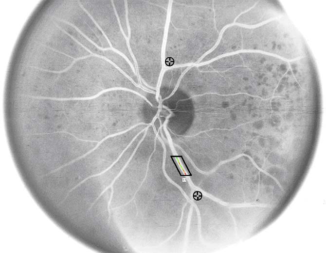 T.I. Metelitsina et al. / Experimental Eye Research 83 (2006) 128e132
correlation from multiple veins from the same eye. SAS 9.1software (Cary, North Carolina, USA) was used for the analy-sis of the data.
T.I. Metelitsina et al. / Experimental Eye Research 83 (2006) 128e132
correlation from multiple veins from the same eye. SAS 9.1software (Cary, North Carolina, USA) was used for the analy-sis of the data.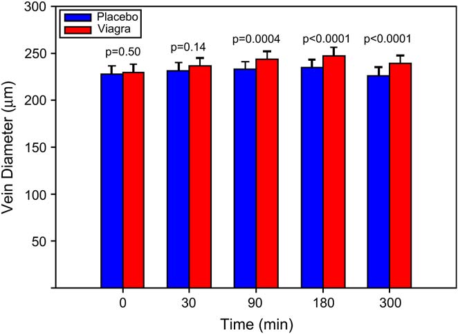 T.I. Metelitsina et al. / Experimental Eye Research 83 (2006) 128e132
significant venous dilatation starting at 90 min and the veinsremained dilatated even at 300 min.
T.I. Metelitsina et al. / Experimental Eye Research 83 (2006) 128e132
significant venous dilatation starting at 90 min and the veinsremained dilatated even at 300 min.