La tétracycline, connue sous le nom commercial Sumycin, agit en bloquant la fixation de l’ARNt sur la sous-unité 30S ribosomale, interrompant l’élongation de la chaîne protéique bactérienne. Ce mécanisme confère une activité sur un spectre large, incluant bactéries Gram positives, Gram négatives, rickettsies et spirochètes. Sa biodisponibilité digestive varie selon la prise alimentaire et les interactions avec les ions divalents comme calcium et magnésium. Sa diffusion tissulaire est importante, notamment dans les voies respiratoires et génito-urinaires. L’élimination se fait par voie rénale et biliaire. Les effets indésirables incluent photosensibilisation, troubles digestifs et coloration dentaire en cas d’administration précoce. Les guides thérapeutiques mentionnent sumycin prix, en soulignant la nécessité de restreindre son utilisation afin de limiter les résistances acquises.
Skinrep239
Skin Research and Technology 2002: 8: 52–56Copyright C Blackwell Munksgaard 2002Blackwell Munksgaard . Printed in DenmarkSkin Research and Technology Age related changes of human skin investigated with histometric measurements by confocal laser scanning microscopy in vivo
Kirsten Sauermann1,2, Sven Clemann1, Sören Jaspers1, Thilo Gambichler2, Peter Altmeyer2,
Klaus Hoffmann2 and Joackim Ennen1
1Department for Research and Development, Beiersdorf AG, Hamburg, Germany, and 2Department of Dermatology, Ruhr-Universität Bochum, GermanyBackground/aims: The confocal laser scanning microscope Results: The older group of volunteers showed a significant
Vivascope (Lucid, Henrietta) allows skin to be studied in real-
increase in Emin, no significant change in DSC, a significant
time with a resolution of 0.5 mm horizontal and 1.3 mm vertical in
decrease in dermal papillae and in the thickness of the basal
vivo. In this study, we present the results of a comparison
layer, and an increase in Agran compared to the younger group.
between the skin of an older and a younger group of volunteers
Conclusions: Histometric measurements by in vivo confocal
by in vivo histometric measurements.
laser scanning microscopy are a sensitive and non-invasive tool
Methods: To investigate changes caused by age, 13 young (18–
for characterizing and quantifying histological changes of the
25 years) and 13 older (Ͼ 65 years) volunteers were examined.
epidermis and papillary dermis due to ageing.
The following parameters were measured using the Vivascopeat the volar forearm: minimal thickness of the epidermis (Emin),
Key words: Confocal laser scanning microscopy – skin ageing –
size of cells in the granular layer (Agran), thickness of the horny
layer (DSC), thickness of the basal layer (DSB) and number ofdermal papillae per area (PapI). The image analysis programimage tool was used to measure the size of the cells and the
INTHEPASTDECADE,advanceshavebeenmadeinim- study on effects of tretionin on chronologically aged
aging human skin in vivo by confocal laser scan-
skin using ultrastructural analysis by transmission
ning microscopy (1, 2). The confocal laser scanning
electron microscopy and light microscopy, Kligman
microscope system, Vivascope 1000, is a commercially
described keratinocytes of the basal and spinous layer
available instrument that allows skin to be studied in
in aged skin as irregular in size and shape (11).
vivo and in real-time imaging. Numerous studies
The aim of this study was to develop histometric
comparing and identifying structures imaged by con-
parameters to investigate and quantify ageing pro-
ventional histological sectioning and the confocal
cesses on human skin in vivo, non-invasively, using
laser scanning microscope have been conducted by M.
the confocal laser scanning microscope.
Rajahyaksha and S. Gonzales (3–5).
Skin that ages chronologically, or enhanced by en-
Material and Methods
vironmental effects such as UV irradiation (6), showssigns, such as wrinkles, that deepen and align in a
parallel pattern, heterogenecity in pigmentation, and
To investigate changes caused by ageing, sites in the
a dry, scaly aspect (7). When these phenomena are in-
middle of the volar forearm of healthy Volunteers,
vestigated by biopsy and histological sectioning and
skin type I to III, were imaged. The volunteers were
staining, dermal elastosis (8), a thin mean epidermal
assigned to one of two groups: one group of volun-
thickness and a flattened epidermal-dermal junction,
teers of greater than 65 years of age (mean 72.5 years,
can be seen (9). The corneocytes shed from the horny
11 female, 2 male), and a younger group, of 18–25
layer increase in size as a result of a lower prolifer-
years (mean 23.1 years, 11 female, 2 male), each group
ation rate and turn-over of the epidermis (10). In a
consisting of 13 volunteers. All investigations were
Age related changes of human skin
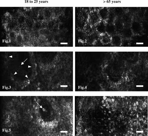
performed in winter. Informed consent was obtainedprior to confocal imaging. InstrumentThe Vivascope 1000 is a confocal laser scanning micro-scope, built by the Lucid Company to examine skinin vivo. The system uses a laser source with a wave-length of 830 nm, an illumination power up to 20 mWon the object, a water immersion objective and an im-aging rate of 20 per second. The field of view shows128 mm ¿ 260 mm. Its lateral resolution is 0.4 mm, thevertical resolution is about 1.9 mm. The microscopeallows imaging of the epidermis and papillary dermison a cellular level. As Millind Rajahyaksha has shownin 1995 (2), melanin provides a strong contrast in theimages of the epidermis, especially between the basaland the spinous layers. Fig. 1. The granular layer appears as large cells with dark nuclei andstrong reflecting cytoplasm containing numerous granules. The size of
All investigations were performed at 21 æC ∫ 1 æC and
cells was measured in the most apical layer of the Str. granulosum
50% ∫ 5% relative humidity. In healthy volunteers, the
(Agran). This picture shows the granular layer of a volunteer aged 23
horny layer of the forearm appears as a relatively ho-
mogeneous, strong, reflective tissue without visible
Fig. 2. In the skin of the older group of volunteers, the cells in the
cellular structures. The thickness of the horny layer
granular layer are bigger in diameter than in the younger group.
(DSC) was measured by focusing through the epider-
Fig. 3. The epidermal-dermal junction appears as bright reflectingcircles of basal layer (melanosomes are strongly reflective) (arrow-
mis and measuring the difference between surface
heads) around darker round areas which represent the dermal papillae
and the depth, at which living cells can be observed
(arrow). Between these circles, spinous layer forming a typical honey-
for the first time, with the micron screw. These cells,
comb-like pattern can be observed. The basal layer in the skin of the
the granular layer of the epidermis, appear as bright
young volunteers appears broad and regular.
circles of intercellular matrix and cytoplasm with nu-
Fig. 4. In the skin of older volunteers, the basal layer shows a more
merous granules surrounding dark nuclei. The meas-
fringed, irregular aspect than in younger volunteers with varying sizes
urement of the DSC was repeated at least five times
Fig. 5. The bright structure (arrow) in the centre of the dermal papillaerepresents blood cells flowing in a capillary. At the epidermal junction,
The size of cells in the granular layer (Agran) was
images were captured and the thickness of the basal layer (arrowhead)
evaluated by taking pictures of the upper granular
was measured by means of an image analysis program.
layer (Figs. 1, 2). In confocal images, this is the most
Fig. 6. Little interdigitation can be seen in aged skin. Large parts of
apical plane of focus showing nucleated cells. The size
the epidermal-dermal junction consist of plane basal layer covering the
of cells was evaluated using an image analysis pro-
dermis. The number of dermal papillae per area decreased compared tothe younger group of volunteers. (scale bars Ω 25 mm)
The minimal thickness of the epidermis (Emin) was
measured, in a similar manner to that of the hornylayer, as the distance between the surface and the
tion at sites where the basal layer was orientated verti-
level showing the first dermal tissue inside the circles
cally compared to the skin surface (Figs. 3, 4) and ana-
representing the basal layer of the epidermis. Eight
lysing them using the image analysis program image
tool. Images of at least 5 dermal papillae were cap-
As a water immersion objective was used, care was
tured. The basal layer of each papilla was measured
taken to ensure that all data relating to the horny layer
and the minimal thickness of the epidermis were ob-
The epidermal-dermal junction of the forearm ap-
tained during the first five minutes of adapting the
pears as bright shining circles of basal layer, contain-
objective and the water immersion to the skin, to
ing melanin and therefore reflecting strongly, with
dark centres, the papillary dermis. These circles are
The thickness of the basal layer (DSB) was evalu-
surrounded by spinous layer cells. The lumen of capil-
ated by taking pictures of the epidermal-dermal junc-
lary loops can be seen in the centre of the dermal pap-
Sauermann et al.
illae as black holes (Fig. 3), often showing bloodflow,
older group. The difference was not statistically sig-
with bright erythrocytes moving through the capillary
(Fig. 5). The number of dermal papillae per area (PapI)
The minimal thickness of the epidermis was sig-
was evaluated by counting each papilla showing the
nificantly higher in the older group than in the
capillary in each field of view; 20 fields of view were
younger one (mean Emin: 40.0 mm ∫ 5.6 mm vs. 35.5 mm
checked. As the dermal papilla structure is big com-
pared to the field of view, it often had to be decided
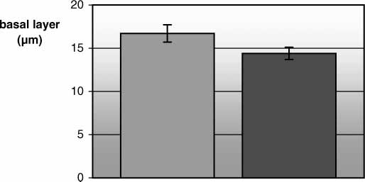
The basal layer in the older skin was significantly
whether or not a papilla should be counted, as only
thinner than that of the younger volunteers and the
parts of it could be seen. Therefore, as there is nor-mally only one small capillary loop in the centre ofthe papillae in the body region investigated, (Fig. 5),the presence of the capillary in the area of view wastaken as the criteria for counting the papillae. (Thisparameter was measured in only 20 out of the 26 vol-unteers.)
Image analysisThe captured images of the skin at the height of theepidermal-dermal junction and at the height of theapical parts of the granular layer were analysed using
Fig. 7. The distance between surface and the top of the dermal papillae,
the image analysis program IMAGE TOOL (UTHS-
the minimal thickness of the epidermis, increases with age. Light
CA, San Antonio, USA). The program was calibrated
grey Ω aged 18 to 25 years, dark grey Ω aged Ͼ65 years.
using a sputtered scale, imaged with the same instru-ment that was used to examine the skin. The thicknessof the basal layer was measured as the thickness ofthe circle of bright reflecting cells (Fig. 5). Five meas-urements per papilla, with at least 25 measurementsper volunteer were performed.
The size of cells in the granular layer was measured
with the same program. As skin is wrinkled, the planeof focus and the granular layer were often not parallelthrough the whole field of view. Sometimes areasshowing deeper layers were apparent in imagesshowing mainly the granular layer. As the size of thekeratinocytes decreases from the granular to the basal
Fig. 8. The thickness of the basal layer measured by image analysis
layer due to the different polarity of the cells, care has
decreases with age. Light grey Ω aged 18 to 25 years, dark grey Ωaged Ͼ65 years.
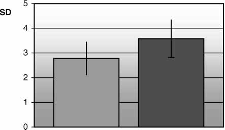
to be taken not to measure spinous cells instead ofcells of the granular layer by mistake. To avoid this,only cells with a broad cytoplasm containing granuleswere chosen and measured. At least 20 cells per areawere measured. Statistical analysisMorphometric results from confocal imaging are ex-pressed as mean ∫SD. A two-tailed student’s t-testwas used to compare the two groups of volunteers. P-values of less than 0.05 were considered significant. Fig. 9. The standard deviation of the single measurements of the thick-ness of the basal layer in each individual is significantly bigger in theolder volunteers, reflecting the irregular aspect of the basal layer ob-
The thickness of the horny layer was 10.4 mm ∫ 3.2 mm
served in their skin. Light grey Ω aged 18 to 25 years, dark grey Ω
in the younger group and 11.2 mm ∫ 1.9 mm in the
Age related changes of human skin Discussion
The thickness of the horny layer did not show anychanges in our study, confirming similar findings inprevious studies (Kligman 1986, Grove 1983).
The changes in minimal thickness of the epidermis
were considered to be due in part to changes in theliving tissue of the epidermis. In previous studies(Kligman 1986), mean thickness of the epidermis de-creased with age. Although this seems to contradict
Fig. 10. The size of cells in the granular layer is bigger in the skin of
our results, these studies also describe the height of
the older group of volunteers compared to the young group, similar to
the epidermal-dermal junction decreasing with age. the known increase with age in the size of corneocytes. Light grey Ωaged 18 to 25 years, dark grey Ω aged Ͼ65 years.
Our results for thickness of the epidermis, immedi-ately above the dermal papillae, could still be consist-ent with a decrease in the mean epidermal thicknessfound by histological methods, if the decrease inheight of the epidermal–dermal junction was morethan twice the decrease in the mean epidermal thick-ness.
The correlation between size of cells in the granular
layer and age is consistent with a documented in-crease in corneocytes with age. We propose that thesize of corneocytes is determined by the size of cellsin the upper granular layer and that comparable re-sults can be obtained using both investigation
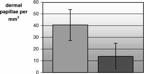
Fig. 11. The number of dermal papillae per area decreases dramatically
methods, providing the kinetics are excluded. with age. Light grey Ω aged 18 to 25 years, dark grey Ω aged Ͼ65
Changes in the thickness of the basal layer are com-
monly attributed to flattening of the epidermal-der-mal junction, however, this is unlikely to be the casein our study, as this should cause an increase in the
mean standard deviation of the 25 measurements of
thickness of the basal layer, by decreasing the angle
each volunteer was greater in the older than in the
between the direction of the basal layer and the plane
younger group (Figs. 8, 9). This reflects the irregular,
of focus and increasing its projection, while the thick-
fringed aspect of the basal layer in aged skin (Fig.
ness of the basal layer in our study was increased. In
4). The mean thickness of the basal layer in the
spite of this, we suggest that this decrease in height
aged group of volunteers was 14.4 mm ∫ 0.7 mm, com-
and increase in irregularity reflects the decrease in
pared to 16.7 mm ∫ 1.0 mm in the younger group, and
proliferation of the basal layer in aged skin. Kligman,
the mean standard deviation of the thickness of the
1993, pointed out that the basal and spinous cells
basal layer in the data of each volunteer was 2.78
showed an irregular shape and size in electron micro-
mm ∫ 0.66 mm in the younger group and 3.58 mm ∫
scopic images when investigating the effects of topical
retinoic acid on chronologically aged skin. As the rate
The cells in the upper granular layer (Agran) were
of proliferation is decreased in aged skin (Grove et al.,
significantly larger in the older group of volunteers:
1983), the irregular shape could be a result of a re-
1004 mm2 ∫ 120 mm2 compared to 803 mm2 ∫ 113 mm2
duced number of the basal layer cells participating in
the cell cycle. The variation in thickness of the basal
The number of dermal papillae per area was sig-
layer could possibly be studied in vivo, dynamically
nificantly lower in the aged group than the younger
The decrease in the number of dermal papillae per
In the younger skin, mean papillae containing capil-
area (PapI) with age reflects the flattening of the epi-
laries was 40.56 ∫ 13.19 per mm2 compared to only
dermal-dermal junction (9). In particular, this demon-
13.82 ∫ 11.11 per mm2 in the aged skin. In the older
strates that not only the height of the dermal papillae
group of volunteers, regions of totally plane basal
decreases with age, but also the number of interdigi-
layer often filled the whole area of view (Fig. 6). Sauermann et al.
The most dramatic age-related changes in this
dermatitis: correlation of in vivo confocal imaging to routine
study were seen in the number of papillae per area.
histology. J Am Acad Dermatol 1999: 40: 708–713.
5. Gonzalez S, Rubinstein G, Mordovtseva V, et al. In vivo ab-
As this parameter was closely linked to its function of
normal keratinazation in Darier-White’s disease as viewed
supplying the epidermis with water and nutrients via
by real-time confocal imaging. J Cutan Pathol 1999: 26: 504–
the dermal vasculature, it seemed to be a more sensi-
6. Kligman LH. Photoaging. Manifestations, prevention and
tive measure for qualitative evaluation of the epider-
treatment (Review). Dermatol Clin 1986: 4: 517–528.
mal junction, than the measurement of height in histo-
7. Kligman LH, Kligman AM. The nature of photoaging: its
logical sections. It can also be obtained easily, quick
prevention and repair. Photodermatol 1986: 3: 215–227.
8. Miyachi Y, Ishikawa O. Dermal connective tissue met-
abolism in photoageing. Australas J Dermatol 1998: 39: 19–
In conclusion, histometric measurements by in vivo
confocal laser scanning microscopy are a sensitive and
9. Lavker RM, Zheng PS, Dong G. Aged skin: a study by light,
non-invasive tool to characterize and quantify histo-
transmission electron and scanning electron microscopy. J
logical changes of the epidermis and papillary dermis
Invest Dematol 1987: 88: suppl. (3): 44–51.
10. Grove GL, Kligman AM. Age-associated changes in human
epidermal cell renewal. J Gerontol 1983: 38: 137–142.
11. Kligman AM, Dogadkina D, Lavker RM. Effects of topical
tretinoin on non-sun-exposed protected skin of the elderly. References
J Am Acad Dermatol 1993: 29: 25–33.
1. Imbert D, Hoogstraate J, Marttin E, Cullander C. Imaging
thick tissues with confocal microscopy. Methods Mol Biol
2. Pierard GE. In vivo confocal microscopy: a new paradigm
in dermatology. Dermatology 1993: 186: 4–5. Department for Research and Development
3. Rajadhyaksha M, Gonzalez S, Zavislan JM, et al. In vivo
confocal scanning laser microscopy of human skin II: ad-
vances in instrumentation and comparison with histology. J
Invest Dermatol 1999: 113: 293–303.
4. Gonzalez S, Gonzalez E, White WM, et al. Allergic contact
e-mail: K.Sauermann/Hamburg.Beiersdorf.com
GLOBALIZATION: WHAT IS NEW; EFFECTIVE GLOBAL Marketing Department, Bloomsburg University, Pennsylvania, Distinguished Professor Emeritus of Marketing and International Business, Concordia University, Montreal, and Distinguished Visiting Professor International Business, Helsinki Professor of International Business, Helsinki School of Economics Abstract What is new and important about glob
Infectious Diseases Society of America Guidelinesfor the Diagnosis and Treatment of AsymptomaticBacteriuria in Adults Lindsay E. Nicolle,1 Suzanne Bradley,2 Richard Colgan,3 James C. Rice,4 Anthony Schaeffer,5 and Thomas M. Hooton6 1University of Manitoba, Winnipeg, Canada; 2University of Michigan, Ann Arbor; 3University of Maryland, Baltimore; 4University of Texas,Galveston; 5Northwestern Univ
 Age related changes of human skin
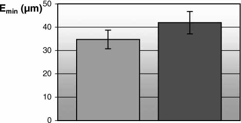
 Sauermann et al.
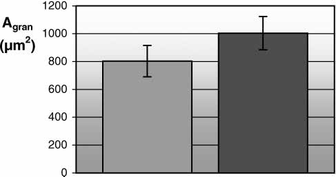
 Age related changes of human skin