La tétracycline, connue sous le nom commercial Sumycin, agit en bloquant la fixation de l’ARNt sur la sous-unité 30S ribosomale, interrompant l’élongation de la chaîne protéique bactérienne. Ce mécanisme confère une activité sur un spectre large, incluant bactéries Gram positives, Gram négatives, rickettsies et spirochètes. Sa biodisponibilité digestive varie selon la prise alimentaire et les interactions avec les ions divalents comme calcium et magnésium. Sa diffusion tissulaire est importante, notamment dans les voies respiratoires et génito-urinaires. L’élimination se fait par voie rénale et biliaire. Les effets indésirables incluent photosensibilisation, troubles digestifs et coloration dentaire en cas d’administration précoce. Les guides thérapeutiques mentionnent sumycin prix, en soulignant la nécessité de restreindre son utilisation afin de limiter les résistances acquises.
Proposed treatment for vestibular dysfunction in dogs
Proposed Treatment for Vestibular Dysfunction in Dogs
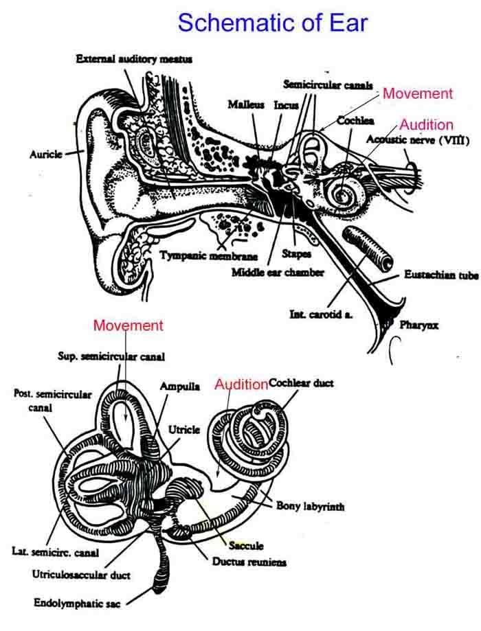
Vestibular dysfunction in the dog can be a disturbing condition for owners, as well as somewhat confounding for the veterinarian and/or physiotherapist. This condition often presents with a sudden onset, no obvious triggering incident, and a very dramatic presentation. The vestibular system is responsible for maintaining the orientation of the animal with respect to its environment. A disruption to the vestibular system results in: ataxia, un-coordination, circling, falling, head tilt (usually in the direction of the lesion), nystagmus (involuntary eye movement), and/or vomiting. The anatomy of the vestibular apparatus in the dog is very similar to that of the human. The inner ear is located in the temporal bone and is comprised of three parts: the vestibule, the cochlea and three semicircular canals. The three semicircular canals are the anterior, posterior, and lateral canals, each positioned at approximately a 90 degree angle in relation to the other two. The three semicircular canals cover the full 360 degrees of movement. The orientation of the canals is also very similar in dogs and humans. The inner ear is filled with fluid and contains two organs: the utricle and the saccule. These organs are receptors, which register tilt and acceleration. In the veterinary literature, vestibular dysfunction is frequently described as “idiopathic peripheral vestibular disease”. Although the clinical signs can be severe, the literature states that patients usually improve within 1-2 weeks. A head tilt, however, may persist. Typical veterinary treatments are often limited to antihistamines (such as diphenhydramine) to decrease anxiety and anorexia, as well as to alleviate the severity of the head tilt and nystagmus. Vestibular disease or dysfunction is a condition that occurs more frequently in older dogs. When lying down, the canine patient often prefers to lie on the affected side and/or may circle toward that side. Dogs with bilateral peripheral vestibular dysfunction may stand with a wide base with their head held close to the ground and may swing their head from side to side. Nystagmus is a frequently occurring sign in cases with vestibular disease. Nystagmus is a reflex eye movement with a slow phase and a fast phase that presents as a result of stimulation or movement of the inner ear.
In humans, distorted vestibular function has been termed BPPV (Benign Paroxysmal Positional Vertigo). Two types of this condition have been identified: primary/idiopathic and secondary/spontaneous. Frequent remission and spontaneous recurrence is most common in primary or idiopathic BPPV, while secondary or symptomatic BPPV can be due to viral infection or can occur from head trauma, such as during sports or motor vehicle accidents. Although spontaneous remission does occur, appropriate intervention will shorten the duration of these episodes. Debris from the utricle can move into any of the canals, resulting in distorted function of the vestibular apparatus and frequently causing severe signs and symptoms similar to those described in dogs. The most common canal for debris to travel is into the posterior canal. The first documentation of crystals in the ear canals was identified on autopsy in humans in 1992. The same presence or finding has not been investigated or identified in dogs. However, we can speculate that the same pathology occurs considering the anatomy is so similar. In an attempt to devise a treatment regime for our canine clients, it would be of value to recognize the similarity of the inner ear anatomy between humans and dogs and to examine the testing and treatment protocols used in humans. As we assess and treat vestibular conditions, it will be important to recognize the characteristics of nystagmus (involuntary eye movement) as the affected canals are placed in a position that causes the signs and symptoms such as vertigo. It should be noted that vertigo is time limited, approximately 30 seconds in duration. There is a delay in the onset of the nystagmus, and it is a response that decreases in intensity each time it is tested for. There are two typical diagnostic tests used in humans to test for debris in both the anterior and posterior canals.
1. The Dix-Hallpike maneuver: is one in which the patient is sits, with legs straight
out in front and with their head turned 45 degrees to the side being tested. The patient is then rapidly lowered into a horizontal position and observed for signs of nystagmus. The patient is then returned to a sitting position with the head still in 45 degrees of rotation and observed for nystagmus again
2. The side lying maneuver: is one in which the patient sits on the edge of the bed
with the head turned 45 degrees away from the side being tested. The patient is then lowered on to their side moving in the direction of the ear being tested, and we note if nystagmus occurs. The patient is then returned to a sitting position with the head still turned away. The patient is observed for nystagmus again.
The above tests will identify which side has the affected canal. In actual practice, the above procedures are usually modified so that once the affected side has been identified,
the practitioner will move directly to the treatment position without returning the patient to sitting. If nystagmus is present when testing both sides, the problem side usually demonstrates the most intense nystagmus. If nystagmus is purely a vertical nystagmus (up / down involuntary eye movement), this is often indicative of a central nervous system (CNS) condition In treating human BPPV, there are several positioning techniques reported to be successful, depending on the canal affected and on the characteristics of the nystagmus. Adapting some of these postural therapeutic techniques for the treatment of the canine patient can be challenging since this requires cooperation on the part of the dog.
1. Semont Liberatory Maneuver: the human patient sits with their head turned away
from the affected side; then the patient is moved to side-lying on the affected side. Once nystagmus subsides, the patient is moved to side-lying on the opposite side while maintaining the head position away from the affected side. A possible alternative for the dog would be to start with the patient on their stomach and then follow the same procedure described above.
2. Horizontal Canal Repositioning Maneuver: the human patient is lays on their back
with their head turned toward the affected side. Once cessation of the nystagmus takes place, the head is turned away from the affected side, and the patient is placed laying face down. Continue movements in 90 degree increments, each time waiting for cessation of the nystagmus before moving to the next position. A similar maneuver could be performed with the canine patient
Even though it would be challenging to adopt these positional maneuvers for treatment of vestibular disease in dogs, these patients are often so severely debilitated by this condition that it would be relatively easy to move them through these positioning changes. Depending on the size of the dog, it would be quite useful for the therapist to be seated with their legs stretched out in front of them on the floor. The dog would then be cradled between their legs allowing the therapist to help control the positioning of the dog and at the same time, the position of the head. To aid in positioning for some of these techniques, a treatment mat (~ 2” thick) can be useful in order to obtain some degree of extension of the head. If the horizontal canal has been identified as being the affected canal, the owner could be encouraged to keep the dog lying on the unaffected side as much as possible for the first 12 hours post treatment. Due to the fact that persistent head tilt is often seen following acute episodes of vestibular dysfunction in dogs, it would be important to evaluate and treat any possible spinal pain (especially at or between cervical vertebrae 1 and 2). Craniosacral techniques would be another useful approach to use in these patients as the acute symptoms resolve. Physiotherapy treatments are very effective in the treatment of Benign Paroxysmal Positional Vertigo, a very distressing and debilitating condition in humans. We can
speculate that similar techniques and treatments could be applied in the canine patient in order to achieve similar results.
Date of Birth: It is very important to complete ALL sections. This form will help you prepare for the initial assessment as trying to remember past episodes and treatments during the actual interview can be difficult. In turn, it helps me understand your illness as the medical chart is not enough. If necessary, your Counsellor is available to help you complete the form. 1) Your exp
La notation sur internet touche aussi les médicaments Mots clés : Médicaments, Notation, Site Participatif, Afssaps Par Pauline Fréour 16/12/2010 | Mise à jour : 19:09 Réagir Crédits photo : François BOUCHON/Le Figaro Depuis un mois, meamedica.fr propose aux internautes de noter leurs médicaments. Une démarche qui n'inquiète pas trop les professionnels. En pleine affaire du Me
 In humans, distorted vestibular function has been termed BPPV (Benign Paroxysmal Positional Vertigo). Two types of this condition have been identified: primary/idiopathic and secondary/spontaneous. Frequent remission and spontaneous recurrence is most common in primary or idiopathic BPPV, while secondary or symptomatic BPPV can be due to viral infection or can occur from head trauma, such as during sports or motor vehicle accidents. Although spontaneous remission does occur, appropriate intervention will shorten the duration of these episodes. Debris from the utricle can move into any of the canals, resulting in distorted function of the vestibular apparatus and frequently causing severe signs and symptoms similar to those described in dogs. The most common canal for debris to travel is into the posterior canal. The first documentation of crystals in the ear canals was identified on autopsy in humans in 1992. The same presence or finding has not been investigated or identified in dogs. However, we can speculate that the same pathology occurs considering the anatomy is so similar. In an attempt to devise a treatment regime for our canine clients, it would be of value to recognize the similarity of the inner ear anatomy between humans and dogs and to examine the testing and treatment protocols used in humans. As we assess and treat vestibular conditions, it will be important to recognize the characteristics of nystagmus (involuntary eye movement) as the affected canals are placed in a position that causes the signs and symptoms such as vertigo. It should be noted that vertigo is time limited, approximately 30 seconds in duration. There is a delay in the onset of the nystagmus, and it is a response that decreases in intensity each time it is tested for. There are two typical diagnostic tests used in humans to test for debris in both the anterior and posterior canals.
1. The Dix-Hallpike maneuver: is one in which the patient is sits, with legs straight
out in front and with their head turned 45 degrees to the side being tested. The patient is then rapidly lowered into a horizontal position and observed for signs of nystagmus. The patient is then returned to a sitting position with the head still in 45 degrees of rotation and observed for nystagmus again
2. The side lying maneuver: is one in which the patient sits on the edge of the bed
with the head turned 45 degrees away from the side being tested. The patient is then lowered on to their side moving in the direction of the ear being tested, and we note if nystagmus occurs. The patient is then returned to a sitting position with the head still turned away. The patient is observed for nystagmus again.
The above tests will identify which side has the affected canal. In actual practice, the above procedures are usually modified so that once the affected side has been identified,
the practitioner will move directly to the treatment position without returning the patient to sitting. If nystagmus is present when testing both sides, the problem side usually demonstrates the most intense nystagmus. If nystagmus is purely a vertical nystagmus (up / down involuntary eye movement), this is often indicative of a central nervous system (CNS) condition In treating human BPPV, there are several positioning techniques reported to be successful, depending on the canal affected and on the characteristics of the nystagmus. Adapting some of these postural therapeutic techniques for the treatment of the canine patient can be challenging since this requires cooperation on the part of the dog.
1. Semont Liberatory Maneuver: the human patient sits with their head turned away
from the affected side; then the patient is moved to side-lying on the affected side. Once nystagmus subsides, the patient is moved to side-lying on the opposite side while maintaining the head position away from the affected side. A possible alternative for the dog would be to start with the patient on their stomach and then follow the same procedure described above.
2. Horizontal Canal Repositioning Maneuver: the human patient is lays on their back
with their head turned toward the affected side. Once cessation of the nystagmus takes place, the head is turned away from the affected side, and the patient is placed laying face down. Continue movements in 90 degree increments, each time waiting for cessation of the nystagmus before moving to the next position. A similar maneuver could be performed with the canine patient
Even though it would be challenging to adopt these positional maneuvers for treatment of vestibular disease in dogs, these patients are often so severely debilitated by this condition that it would be relatively easy to move them through these positioning changes. Depending on the size of the dog, it would be quite useful for the therapist to be seated with their legs stretched out in front of them on the floor. The dog would then be cradled between their legs allowing the therapist to help control the positioning of the dog and at the same time, the position of the head. To aid in positioning for some of these techniques, a treatment mat (~ 2” thick) can be useful in order to obtain some degree of extension of the head. If the horizontal canal has been identified as being the affected canal, the owner could be encouraged to keep the dog lying on the unaffected side as much as possible for the first 12 hours post treatment. Due to the fact that persistent head tilt is often seen following acute episodes of vestibular dysfunction in dogs, it would be important to evaluate and treat any possible spinal pain (especially at or between cervical vertebrae 1 and 2). Craniosacral techniques would be another useful approach to use in these patients as the acute symptoms resolve. Physiotherapy treatments are very effective in the treatment of Benign Paroxysmal Positional Vertigo, a very distressing and debilitating condition in humans. We can
speculate that similar techniques and treatments could be applied in the canine patient in order to achieve similar results.
In humans, distorted vestibular function has been termed BPPV (Benign Paroxysmal Positional Vertigo). Two types of this condition have been identified: primary/idiopathic and secondary/spontaneous. Frequent remission and spontaneous recurrence is most common in primary or idiopathic BPPV, while secondary or symptomatic BPPV can be due to viral infection or can occur from head trauma, such as during sports or motor vehicle accidents. Although spontaneous remission does occur, appropriate intervention will shorten the duration of these episodes. Debris from the utricle can move into any of the canals, resulting in distorted function of the vestibular apparatus and frequently causing severe signs and symptoms similar to those described in dogs. The most common canal for debris to travel is into the posterior canal. The first documentation of crystals in the ear canals was identified on autopsy in humans in 1992. The same presence or finding has not been investigated or identified in dogs. However, we can speculate that the same pathology occurs considering the anatomy is so similar. In an attempt to devise a treatment regime for our canine clients, it would be of value to recognize the similarity of the inner ear anatomy between humans and dogs and to examine the testing and treatment protocols used in humans. As we assess and treat vestibular conditions, it will be important to recognize the characteristics of nystagmus (involuntary eye movement) as the affected canals are placed in a position that causes the signs and symptoms such as vertigo. It should be noted that vertigo is time limited, approximately 30 seconds in duration. There is a delay in the onset of the nystagmus, and it is a response that decreases in intensity each time it is tested for. There are two typical diagnostic tests used in humans to test for debris in both the anterior and posterior canals.
1. The Dix-Hallpike maneuver: is one in which the patient is sits, with legs straight
out in front and with their head turned 45 degrees to the side being tested. The patient is then rapidly lowered into a horizontal position and observed for signs of nystagmus. The patient is then returned to a sitting position with the head still in 45 degrees of rotation and observed for nystagmus again
2. The side lying maneuver: is one in which the patient sits on the edge of the bed
with the head turned 45 degrees away from the side being tested. The patient is then lowered on to their side moving in the direction of the ear being tested, and we note if nystagmus occurs. The patient is then returned to a sitting position with the head still turned away. The patient is observed for nystagmus again.
The above tests will identify which side has the affected canal. In actual practice, the above procedures are usually modified so that once the affected side has been identified,
the practitioner will move directly to the treatment position without returning the patient to sitting. If nystagmus is present when testing both sides, the problem side usually demonstrates the most intense nystagmus. If nystagmus is purely a vertical nystagmus (up / down involuntary eye movement), this is often indicative of a central nervous system (CNS) condition In treating human BPPV, there are several positioning techniques reported to be successful, depending on the canal affected and on the characteristics of the nystagmus. Adapting some of these postural therapeutic techniques for the treatment of the canine patient can be challenging since this requires cooperation on the part of the dog.
1. Semont Liberatory Maneuver: the human patient sits with their head turned away
from the affected side; then the patient is moved to side-lying on the affected side. Once nystagmus subsides, the patient is moved to side-lying on the opposite side while maintaining the head position away from the affected side. A possible alternative for the dog would be to start with the patient on their stomach and then follow the same procedure described above.
2. Horizontal Canal Repositioning Maneuver: the human patient is lays on their back
with their head turned toward the affected side. Once cessation of the nystagmus takes place, the head is turned away from the affected side, and the patient is placed laying face down. Continue movements in 90 degree increments, each time waiting for cessation of the nystagmus before moving to the next position. A similar maneuver could be performed with the canine patient
Even though it would be challenging to adopt these positional maneuvers for treatment of vestibular disease in dogs, these patients are often so severely debilitated by this condition that it would be relatively easy to move them through these positioning changes. Depending on the size of the dog, it would be quite useful for the therapist to be seated with their legs stretched out in front of them on the floor. The dog would then be cradled between their legs allowing the therapist to help control the positioning of the dog and at the same time, the position of the head. To aid in positioning for some of these techniques, a treatment mat (~ 2” thick) can be useful in order to obtain some degree of extension of the head. If the horizontal canal has been identified as being the affected canal, the owner could be encouraged to keep the dog lying on the unaffected side as much as possible for the first 12 hours post treatment. Due to the fact that persistent head tilt is often seen following acute episodes of vestibular dysfunction in dogs, it would be important to evaluate and treat any possible spinal pain (especially at or between cervical vertebrae 1 and 2). Craniosacral techniques would be another useful approach to use in these patients as the acute symptoms resolve. Physiotherapy treatments are very effective in the treatment of Benign Paroxysmal Positional Vertigo, a very distressing and debilitating condition in humans. We can
speculate that similar techniques and treatments could be applied in the canine patient in order to achieve similar results.