ISSN 0003-6838, Applied Biochemistry and Microbiology, 2006, Vol. 42, No. 6, pp. 625ŌĆō630. MAIK ŌĆ£Nauka /InterperiodicaŌĆØ (Russia), 2006. Original Russian Text A.P. Bonartsev, G.A. Bonartseva, T.K. Makhina, V.L. Myshkina, E.S. Luchinina, V.A. Livshits, A.P. Boskhomdzhiev, V.S. Markin, A.L. Iordanskii, 2006,published in Prikladnaya Biokhimiya i Mikrobiologiya, 2006, Vol. 42, No. 6, pp. 710ŌĆō715.New Poly-(3-hydroxybutyrate)-Based Systems for Controlled Release of Dipyridamole and Indomethacin A. P. Bonartsevc, G. A. Bonartsevaa, T. K. Makhinaa, V. L. Myshkinaa, E. S. Luchininaa, V. A. Livshitsa, A. P. Boskhomdzhieva, V. S. Markinb, and A. L. Iordanskiic a Bach Institute of Biochemistry, Russian Academy of Sciences, Moscow, 119071 Russiab Semenov Institute of Chemical Physics, Russian Academy of Sciences, Moscow, 119991 Russiac Faculty of Biology, Moscow State University, Moscow, 119899AbstractŌĆöNew poly-(3-hydroxybutyrate)-based systems for controlled release of anti-inflammatory and anti- thrombogenic drugs have been studied. The release occurs via two mechanisms (diffusion and degradation) operating simultaneously. Dipyridamole and indomethacin diffusion processes determine the rate of the release at the early stages of the contact of the system with the environment (the first 6ŌĆō8 h). The coefficient of the release diffusion of a drug depends on its nature, the thickness of the poly-(3-hydroxybutyrate) films containing the drug, the concentrations of dipyridamole and indomethacin, and the molecular weight of the poly-(3- hydroxybutyrate). The results obtained are critical for developing systems of release of diverse drugs, thus, enabling the attainment of the requisite physiological effects on tissues and organs of humans. DOI: 10.1134/S0003683806060159
Poly-(-3-hydroxybutyrate) (PHB) and its copoly-
enclosing the implant and, if the polymer contacts vas-
mers obtained using biotechnological methods have
cular tissues, the extent of the hypertrophy of the blood
become the subject of increasing interest due to their
vessel walls. As is clear from the above, the processes
biodegradability and biocompatibility, which make it
constituting the organismŌĆÖs response to implantation of
possible to use these polymers in medicine. The physi-
biodegradable polymers (i.e., inflammation, thrombus
cochemical and biological properties of PHB allow this
formation, and cell proliferation) need to be regulated
polymer to be used as a material for implantable medi-
cal devices (e.g., membranes for treatment of periodon-
The buildup of inflammatory and thrombogenic pro-
tal disease and prevention of adhesions) and coatings
cesses may be regulated by systemic administration of
(to be applied onto the surface of net endoprostheses,
antiaggregant and anti-inflammatory preparations. In
pacemakers, stents, vascular prostheses, etc.) [1].
certain cases, however, this approach is not efficient,
Implantation of devices made from biodegradable
because the local concentrations of the drugs within the
materials, including PHB and its copolymers, into tis-
region of the implantation are either not sufficient for
sues of an organism may be associated with a series of
attaining the pharmacological effect or lack stability,
undesirable processes; these include pathological
whereas any further increase in the dose administered
inflammatory reactions, formation of thrombi, and the
systemically entails side effects [3].
lack of correspondence between the rates of the implant
Systems of controlled release of drugs, based on
replacement by the surrounding body tissues and the
polymer materials, make it possible to regulate the pro-
rate of its biodegradation (which may be higher or
cesses of inflammation, thrombus formation, and devel-
lower). The character of the inflammatory process
opment of new tissue within the immediate vicinity of
accompanying polymer implantation determines to a
implantation of medical devices. In designing such sys-
considerable extent the intensity of the biodegradation
tems, it is important to make the right choice of the
of this polymer. The success of the integration of an
drug. Dipyridamole (DP), a widely used antithrombo-
implant into the surrounding tissues (if there is a con-
genic drug, is a phosphodiesterase inhibitor promoting
tact between the implant and blood or intraperitoneal
intracellular accumulation of cGMP and cAMP, which
fluid) depends in its resistance to thrombus formation.
inhibits both platelet aggregation and cell proliferation
The increased coagulability of peritoneal fluid favors
[4]. Indomethacin (IM), a nonsteroidal anti-inflamma-
the development of peritoneal adhesions, which is a
tory drug (NSAID), inhibits cyclooxygenase, thereby
serious pathology. The intensity of the cell proliferation
preventing the synthesis of prostaglandins (which are
associated with polymer implantation determines both
major mediators of inflammation), and cell prolifera-
the rate of formation of a connective tissue capsule
tion [5]. It is noteworthy that DP and IM, as well as
PHB, are soluble in organic solvents (chloroform and
added to 2 ml of the suspension, and the mixture was
methylene chloride), which simplifies the technology
heated at 100°ë for 2 h (water bath); the insoluble res-
of creating polymer systems of controlled release.
idue (PHB granules) was separated by centrifugation at
The molecular weight (MW) of a polymer consider-
8000 g for 20 min. Following the addition of 5 ml of
ably affects the kinetics of the release of drugs intro-
chloroform to the residue, the tube was hermetically
duced into its matrix [4]; for this reason, development
sealed and incubated overnight (28°ë) under continu-
of controlled release systems for drugs that have pre-
ous shaking (shaker). Thereafter, the tube was centri-
defined characteristics requires a technology for syn-
fuged and the chloroform extract was dried in an air
thesizing polymers with a particular MW. When PHB is
flow. Following the addition of concentrated sulfuric
obtained using biotechnological methods, the condi-
acid (5 ml per each 0.1 ml of extract), the mixture was
tions of culturing of the PHB producer strains may
heated at 100°ë for 10 min (water bath) and allowed to
influence the molecular weight of the polymer [6].
cool. The amount of crotonic acid (formed as a result of
Thus, a technology for the biosynthesis of PHB with a
acidic hydrolysis of PHB and subsequent hydroxybu-
defined MW is prerequisite to creating controlled
tyrate dehydration) was measured at 235 nm (against
release systems for the requisite characteristics of the
concentrated sulfuric acid) on a Beckman DU-650
kinetics of the drug release from the polymer matrix.
spectrophotometer (Germany) in 1-cm cuvettes [8].
The use of such systems for controlled release of
The MW of the polymer was determined viscomet-
antithrombogenic and anti-inflammatory drugs is
rically. Measurement of the changes in the viscosity of
expected to (a) increase the resistance of medical
the PHB solution in chloroform were performed at
devices contacting blood (e.g., coatings of stents and
30┬░C. The MW was calculated using the MarkŌĆōHou-
vascular prostheses) to thrombus formation, (b) regu-
winkŌĆōKuhn equation; the value of the coefficient [╬Ę]
late inflammatory processes and the rate of the implant
biodegradation and capsulation (e.g., in the case of
The chemical structure of the polymer, the type of
reticular endoprostheses for hernioplasty and mem-
its crystal lattice, and the extent of its crystallinity
branes for treatment of periodontal disease), and
(0.74) were previously determined using the methods
(c) prevent the formation of adhesions (endoprostheses
of differential scanning calorimetry, IR Fourier spec-
for hernioplasty and anti-adhesion membranes).
troscopy, and crystal X-ray structure analysis [10].
In this work, we sought to obtain and study PHB-
The traces of residual solvent were controlled by
based films incorporating DP and IM.
measuring the IR spectra on a Brucker IFS-48 IR spec-trometer (Germany). The extent of the weight lossresulting from degradation was determined gravimetri-
The PHB producer strain used in this work (Azoto-
In experiments aimed at studying the kinetic charac-
bacter chroococcum 7B) was capable of synthesizing
teristics of the drug release from the PHB matrix, two
PHB in an amount of up to 80% of the dry weight of the
PHB batches were used differing in their MWs:
bacterial cells. The strains were isolated from the rhizo-
320 kDa (low-molecular-weight PHB) and 1470 kDa
sphere of wheat (sod-podzol soil). A collection of
(high-molecular-weight PHB). The PHB films were 10,
strains of the genus Azotobacter were maintained on
20, or 40 ┬Ąm thick, containing 3.3, 10, or 30 wt %,
AshbeyŌĆÖs medium [6]. To achieve cellular PHB hyper-
respectively, of DP or IM. Systems with a predefined
production, the culture of the Azotobacter strain was
content of the drugs were prepared by evaporating chlo-
grown on BurkeŌĆÖs medium under conditions of an
roform on a glass substratum. In addition to films, a
excess content of the source of carbon (g/l):
polypropylene net was studied, which was modified by
├źgSO4 ┬Ę 7H2O, 0.4; FeSO4 ┬Ę 7H2O, 0.01; Na2MoO4 ┬Ę
applying onto its surface a polymer composition con-
2H2O, 0.006; trisodium citrate, 0.5; CaCl2, 0.1;
taining PHB (320 kDa) and DP (10 wt %).
K2HPO4 ┬Ę 3H2O, 1.05; KH2PO4, 0.2; and sucrose, 40 [6, 7].
The rate of the drug release was recorded by UV
The process of isolation and purification of the poly-
spectrometry (DU-650) within the region of maximum
mer from the biomass of A. chroococcum 7B included
absorption of aqueous solutions of DP and IM (at 293
the following stages: dissolution of PHB in chloroform
and 256 nm, respectively). The release was performed
by shaking at 37°ë for 12 h (shaker), separation of the
in phosphate-buffered saline (pH 7.4) at 37°ë for 18 h.
PHB solution from the cell residue by filtration, isola-tion of the PHB by isopropanol precipitation, andrepeated dissolution of the PHB in chloroform followed
by isopropanol precipitation and drying at 60°ë. Effect of the conditions of culturing on the molec-
The content of PHB in the cells was determined
ular weight of the poly-(3-hydroxybutyrate) synthe-
using the method of Zevenhuisen [8]. A suspension of
sized. In experiments addressing the effects of the con-
the cells (20ŌĆō100 mg of dry biomass) was centrifuged
ditions of the culturing on the MW of the polymer syn-
at 5000 g for 20 min. Thereafter, the cells were resus-
thesized, we varied the concentration of the
pended in 10 ml water and homogenized. 2M HCl was
APPLIED BIOCHEMISTRY AND MICROBIOLOGY Vol. 42 No. 6 2006
NEW POLY-(3-HYDROXYBUTYRATE)-BASED SYSTEMS
As demonstrated previously, the addition of organic
Table 1. Effect of a supplemental carbon source (sodium ac-
acids to a sucrose-containing medium decreases the
etate) on the molecular weight (MW) of PHB synthesized by
MW of the polymer synthesized [6]. For this reason, we
designed experiments in which we varied the concen-tration of sodium acetate in the culture medium. The
results obtained are summed up in Table 1. On increas-
ing the concentration of sodium acetate in the mediumfrom 0 to 5 g/l (the content of sucrose, which served asthe primary source of carbon, remained constant and
was equal to 40 g/l), we observed a decrease in theMW of the PHB synthesized by the cells of A. chroo-
It is conceivable that an increase in the intracellular
concentration of the acetyl groups stimulates the activ-ity of acetoacetyl-CoA reductase (EC 1.1.1.36), which,in turn, elevates the content of hydroxybutyryl-CoA. At
high concentrations of acetate, the numbers of poly-merization centers and initial fragments of polymer
* Molecular weights of the PHB batches used for creating systems
chains increase, which results in the synthesis of PHB
for controlled release of dipyridamole and indomethacin.
Thus, the method used in this work makes it possi-
ble to synthesize PHB with a defined MW.
rate of release is near-constant. Our analysis of the
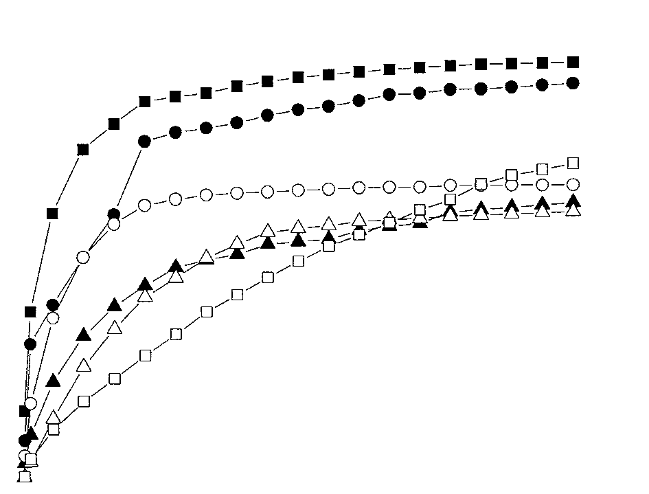
Studies of the kinetics of drug release from a
curves presented in Fig. 1 demonstrates that the mech-
poly-(3-hydroxybutyrate) matrix. Figure 1 shows
anism of release is determined by a superposition of
typical kinetic curves of DP and IM release from PHB
two processes: (1) DP and IM desorption proper (diffu-
films (each graph is a time dependence of the relative
sion mechanism) and (2) hydrolytic PHB degradation
amount (%) of the drug released). As is evident from
(which becomes most obvious when the first, diffusion-
the figure, most of the systems lack constant limiting
related stage has been completed). As a result of this
values of the concentrations, which would be observed
degradation, the release of the drugs is linear over the
if the release were underlain solely by diffusion mech-
anisms. These kinetic curves are characterized by the
To analyze the kinetics of the release, we subtracted
presence of an initial nonlinear (with respect to time)
the linear input of the hydrolytic degradation from the
segment and a terminal linear segment within which the
common current values of the concentration of the drug
Fig. 1. Kinetic curves of drug release: IM 10% (1ŌĆō3) and DP 10% (4, 5) and 30% (1ŌĆō4) from PHB (MW = 320 kDa) Fig. 2. Kinetic desorption curves of indomethacin (1ŌĆō3) and
films with a thickness of 10 ┬Ąm (1ŌĆō4) and 20 ┬Ąm (6) or a
dipyridamole (4ŌĆō6) following the diffusion mechanism.
polypropylene surgical net coated with a 20 ┬Ą layer of PHB
The samples of PHB (MW = 320 kDa) used had a thickness
of 10 (1, 4), 20 (2, 5), and 40 (3, 6) ┬Ąm.
APPLIED BIOCHEMISTRY AND MICROBIOLOGY Vol. 42 No. 6 2006
ues of the freely diffusing component (DP or IM) than
their thicker counterparts. This result may be accounted
for by the observation that thin films preclude organiza-tion of perfect crystalline structures, this being the rea-
son why the sorption capacity of the low-molecular-
weight component in such polymer systems increases
The analysis of the kinetic curves in the diffusion
equation 1 plots makes it possible to calculate the dif-
fusion coefficients of the drugs and, consequently, givea quantitative characterization of the systems for con-
ŌłéG/Ōłét = D[Ōłé2G/Ōłéx2] + k, (1)
where D is the diffusion coefficient (of DP or IM),
cm2/s; k is the constant of the polymer hydrolysis, sŌĆō1;
G is the concentration (of DP or IM), %; and x and t are
Fig. 3. Graphical solution of the diffusion equation for
the coordinate position (cm) and time (s) of the diffu-
determining the coefficient of dipyridamole diffusion in
PHB (MW = 320 kDa) films with a thickness of 10 (1),
The solution of this equation for the condition
20 (2), and 40 (3) ┬Ąm. Gt/Goo > 0.5 has the classic appearance
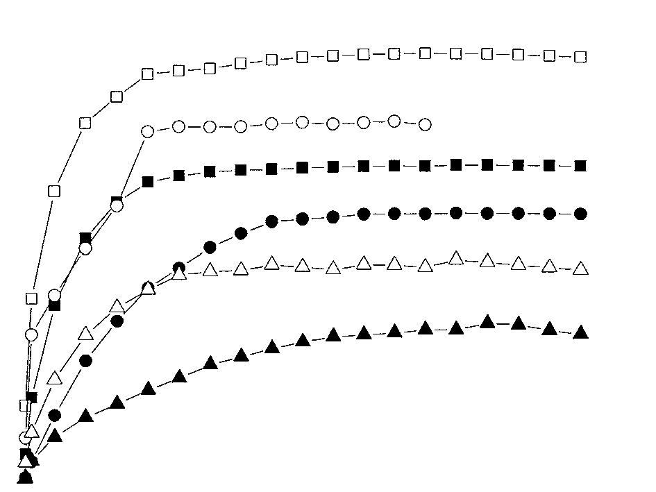
released (such as those shown in Fig. 1). The result of
Gt/Goo = 1 ŌĆō (8/ŽĆ2exp(ŌĆōDt/L2),
this data processing characterizing the diffusion pro-cess proper is depicted in Fig. 2. Figure 2 shows that
where L is the thickness of the PHB film, cm (the other
thin PHB films (10 ┬Ąm thick) have higher limiting val-
designations being the same as in Eq. 1).
If the logarithm of this equation is taken, the diffu-
sion coefficients may be determined by solving the
Table 2. Diffusion parameters of the system PHBŌĆōdrug (DP
graphical equation in log (1 ŌĆō G /G )
Examples of such solutions are shown in Fig. 3 for
DP diffusion from films of variable thickness. The val-
ues of the diffusion coefficient, calculated using equa-tion (3), are listed in Table 2.
Diffusion coefficients are known to characterize the
mobility of polymeric segments, the morphology of
PHB, and the intensity of the interactions of the drugwith functional groups (in this case, ester groups) of the
polymer. The rate of the diffusion-mediated release is
higher for IM than DP, all other conditions (i.e., the filmthickness and drug concentration) being the same. The
maximum sorption capacity of PHB is also higher for
IM than DP, regardless of the film thickness, as Fig. 4demonstrates. It is exactly this amount of the drug that
is contained within PHB in a nonimmobilized form
capable of free diffusion from the matrix. Thus, thenature of the drug considerably affects the rate of its
release, which is particularly important in the case of
combined systems releasing two or more drugs.
The rate of the drug release also depends on the MW
diffusion coefficient of DP was two times greater in the
case of the low-molecular-weight PHB (320 kDa) ascompared to the high-molecular-weight species
(1470 kDa). It is conceivable that the higher rate of the
drug release from the matrix of the low-molecular-
APPLIED BIOCHEMISTRY AND MICROBIOLOGY Vol. 42 No. 6 2006
NEW POLY-(3-HYDROXYBUTYRATE)-BASED SYSTEMS
Fig. 4. Dependence of the maximum concentration of the Fig. 5. Rate of release (R, ┬Ąg/day per cm2) of dipy-
freely diffusing component (Goo) on the thickness (L, ┬Ąm)
ridamole (1, 2) and indomethacin (3) from a matrix of
of a PHB (MW = 320 kDa) film for dipyridamole (1) and
PHB (MW = 320 kDa): 1, 30 wt % DP; 2, 10 wt % DP; and
weight PHB is accounted for by the greater mobility of
PHB films containing the drug, the concentrations of
its polymeric segments. However, the relationship was
DP and IM, and on the MW of the PHB. The results
reversed when we examined 10-┬Ąm films. This obser-
obtained are critical for developing systems of release
vation may be underlain by the fact that the organiza-
of diverse drugs enabling the attainment of the requisite
tion of the polymer molecules in thin films is lower than
physiological effects on tissues and organs of human
In recent years, systems for controlled release of DP
and IM based on other biodegradable polymers (e.g.,
polylactides and copolymers thereof) have been thesubject of active development and investigation [13,
This work was supported in part by state contract
14]. Judging by the reported evidence, these systems
no. 02.467.11.3004 of March 30, 2005, which was con-
are pharmacologically efficient. The results of our stud-
cluded within the framework of an integrated project of
ies (the kinetics of drug release from PHB matrices and
the Federal Targeted Scientific and Technological Pro-
the underlying mechanisms) are comparable with these
gram ŌĆ£Live SystemsŌĆØ for the years 2005ŌĆō2006, and by
data. Moreover, our observation that the release of DP
the Russian Foundation for Basic Research (project
and IM from the PHB matrix occurs at a uniform rate
and for a considerable period of time (Fig. 5) makes itpossible to use these systems for long-term regulationof processes involving inflammation, thrombus forma-
tion, and tissue proliferation in the immediate vicinity
1. Chen, G.-Q. and Wu, Q., Biomaterials, 2005, vol. 26,
of the implantation zone. The possibility to regulate the
rate of drug release from the matrix by changing theMW of the PHB offers an opportunity to design PHB
2. Shtilman, M.I., Polymeric Biomaterials. Part I: Polymer
systems for controlled drug release with predefined
Implants, Utrecht: VSP. Science Press, 1993, pp. 3ŌĆō28.
3. Controlled Drug Delivery: Fundamentals and Applica-
In conclusion, we propose new polymer systems
tions, Robinson, J.R. and Lee, V.H.L., Eds., New York:
(PHB-based) for controlled release of anti-inflamma-
tory and antithrombogenic drugs. The release occurs
4. Aktas, B., Utz, A., Hoenig-Liedl, P., Walter, U., and Gei-
via two mechanisms (diffusion and degradation) oper-
ger, J., Stroke, 2003, vol. 34, pp. 764ŌĆō769.
ating simultaneously. The diffusion of dipyridamoleand indomethacin, which determines the rate of the
5. Fosslien, E., Crit. Rev. Clin. Lab. Sci., 2000, vol. 37,
release at the early stages of contact of the system with
the environment (the first 6ŌĆō8 days), is examined in
6. RF Patent No. 2 194 759, Byull. Izobret., 2001.
detail. The coefficients of diffusion are shown todepend on the nature of the drug, the thickness of the
7. RF Patent No. 2 201 453, Byull. Izobret., 2001.
APPLIED BIOCHEMISTRY AND MICROBIOLOGY Vol. 42 No. 6 2006
8. Zevenhuizen, L.P., Antonie van Leeuwenhoek, 1981,
12. Iordanskii, A.L., Rudakova, T.E., and Zaikov, G.E.,
Interaction of Polymers with Bioactive and Corrosive
9. Akita, S., Einada, Y., Miyaki, Y., and Fugita, H., Macro-Media. Ser. New Concepts in Polymer Science, Utrecht:
mol., 1976, vol. 9, no. 2, pp. 774ŌĆō780.
10. Rebrov, A.V., Dubinskii, V.A., Nekrasov, Yu.P., Bonart-
13. Puebla, P., Pastoriza, P., Barcia, E., and Fernandez-Car-
seva, G.A., Shtamm, M., and Antipov, E.M., Vysokomol.
ballido, A.J., Microencapsul., 2005, vol. 22, no. 7,
Soedin., 2002, vol. 44, no. 2, pp. 347ŌĆō351.
11. Suzuki, T., Deguchi, H., Yamane, T., Shimizu, S., and
14. Zhu, W., Masaki, T., Bae, Y.H., Rathi, R., Cheung, A.K.,
Gekko, K., Appl. Microbiol. Biotechnol., 1988, vol. 27,
and Kern, S.E., J. Biomed. Mater. Res., Ser. B: App. Bio-mater., 2006, vol. 77, no. 1, pp. 135ŌĆō143.
APPLIED BIOCHEMISTRY AND MICROBIOLOGY Vol. 42 No. 6 2006
IN THE HIGH COURT OF NEW ZEALAND AUCKLAND REGISTRY CIV-2009-404-003620 THE HAT FACTORY 2006 LIMITEDFourth DefendantF C Deliu and R Zhao for plaintiffB M Cunningham for defendants RESERVED JUDGMENT OF HUGH WILLIAMS J This judgment was delivered by The Hon. Justice Hugh Williams on pursuant to Rule 11.5 of the High Court Rules ŌĆ”ŌĆ”ŌĆ”ŌĆ”ŌĆ”ŌĆ”ŌĆ”ŌĆ”ŌĆ”ŌĆ”ŌĆ”ŌĆ”ŌĆ”ŌĆ
 NEW POLY-(3-HYDROXYBUTYRATE)-BASED SYSTEMS
As demonstrated previously, the addition of organic
Table 1. Effect of a supplemental carbon source (sodium ac-
NEW POLY-(3-HYDROXYBUTYRATE)-BASED SYSTEMS
As demonstrated previously, the addition of organic
Table 1. Effect of a supplemental carbon source (sodium ac-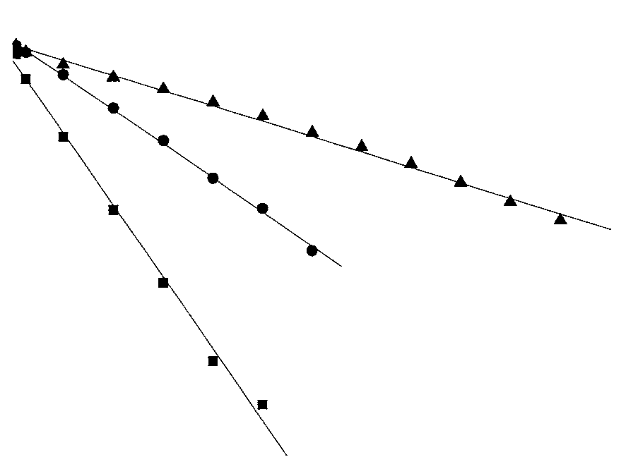 ues of the freely diffusing component (DP or IM) than
their thicker counterparts. This result may be accounted
for by the observation that thin films preclude organiza-tion of perfect crystalline structures, this being the rea-
son why the sorption capacity of the low-molecular-
weight component in such polymer systems increases
The analysis of the kinetic curves in the diffusion
equation 1 plots makes it possible to calculate the dif-
fusion coefficients of the drugs and, consequently, givea quantitative characterization of the systems for con-
ŌłéG/Ōłét = D[Ōłé2G/Ōłéx2] + k, (1)
where D is the diffusion coefficient (of DP or IM),
cm2/s; k is the constant of the polymer hydrolysis, sŌĆō1;
G is the concentration (of DP or IM), %; and x and t are
Fig. 3. Graphical solution of the diffusion equation for
ues of the freely diffusing component (DP or IM) than
their thicker counterparts. This result may be accounted
for by the observation that thin films preclude organiza-tion of perfect crystalline structures, this being the rea-
son why the sorption capacity of the low-molecular-
weight component in such polymer systems increases
The analysis of the kinetic curves in the diffusion
equation 1 plots makes it possible to calculate the dif-
fusion coefficients of the drugs and, consequently, givea quantitative characterization of the systems for con-
ŌłéG/Ōłét = D[Ōłé2G/Ōłéx2] + k, (1)
where D is the diffusion coefficient (of DP or IM),
cm2/s; k is the constant of the polymer hydrolysis, sŌĆō1;
G is the concentration (of DP or IM), %; and x and t are
Fig. 3. Graphical solution of the diffusion equation for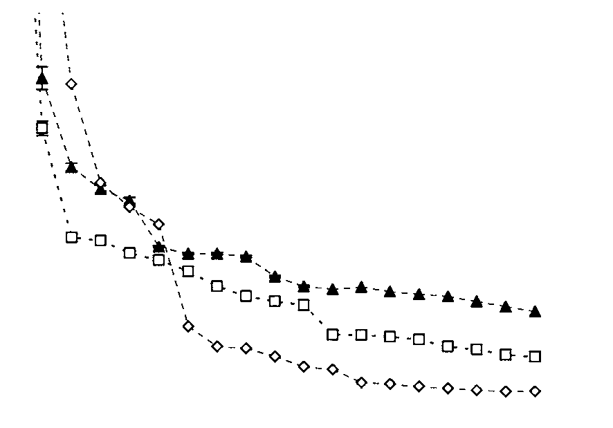 NEW POLY-(3-HYDROXYBUTYRATE)-BASED SYSTEMS
Fig. 4. Dependence of the maximum concentration of the
NEW POLY-(3-HYDROXYBUTYRATE)-BASED SYSTEMS
Fig. 4. Dependence of the maximum concentration of the