La tétracycline, connue sous le nom commercial Sumycin, agit en bloquant la fixation de l’ARNt sur la sous-unité 30S ribosomale, interrompant l’élongation de la chaîne protéique bactérienne. Ce mécanisme confère une activité sur un spectre large, incluant bactéries Gram positives, Gram négatives, rickettsies et spirochètes. Sa biodisponibilité digestive varie selon la prise alimentaire et les interactions avec les ions divalents comme calcium et magnésium. Sa diffusion tissulaire est importante, notamment dans les voies respiratoires et génito-urinaires. L’élimination se fait par voie rénale et biliaire. Les effets indésirables incluent photosensibilisation, troubles digestifs et coloration dentaire en cas d’administration précoce. Les guides thérapeutiques mentionnent sumycin prix, en soulignant la nécessité de restreindre son utilisation afin de limiter les résistances acquises.
Biology.utah.edu
Am. J. Trop. Med. Hyg., 70(2), 2004, pp. 119–124
Copyright 2004 by The American Society of Tropical Medicine and Hygiene
A NOVEL DNA-BASED MICROFLUORIMETRIC METHOD TO EVALUATE
YOLANDA CORBETT, LIURIS HERRERA, JOSE GONZALEZ, LUIS CUBILLA, TODD L. CAPSON,
PHYLLIS D. COLEY, THOMAS A. KURSAR, LUZ I. ROMERO, AND EDUARDO ORTEGA-BARRIA
Instituto de Investigaciones Científicas Avanzadas y Servicios de Alta Tecnología, Ciudad del Saber, Clayton, Panama; Laboratoriode Productos Naturales, Universidad de Panama, Panama City, Panama; Smithsonian Tropical Research Institute, Ancon, Panama;Department of Biology, University of Utah, Salt Lake City, UtahAbstract. This paper describes the development of a novel microfluorimetric assay to measure the inhibition of
Plasmodium falciparum based on the detection of parasitic DNA by intercalation with PicoGreen®. The method was
used to determine parasite inhibition profiles and 50% inhibitory concentration values of known or potential anti-
malarial drugs. Values for parasite inhibition with known anti-malarial drugs using the PicoGreen® assay were compa-
rable with those determined by the standard method based upon the uptake of 3H-hypoxanthine and the Giemsa stain
microscopic technique. The PicoGreen® assay is rapid, sensitive, reproducible, easily interpreted, and ideally suited for
screening of large numbers of samples for anti-malarial drug development.
chrome PicoGreen® into Plasmodium DNA. PicoGreen® is
an ultrasensitive fluorescent nucleic acid stain for measuring
Malaria is among the most life-threatening and widespread
double-stranded DNA (dsDNA) in solution, and it enables
diseases in the world, causing 250−300 million cases and ap-
the detection of quantities as low as 25 pg/mL of dsDNA with
proximately two million deaths annually.1 The disease is
a moderately priced spectrofluorometer using fluorescein ex-
caused by four Plasmodium species (i.e., P. falciparum, P.
citation and emission wavelengths. Accordingly, the micro-
vivax, P. ovale, and P. malariae) that are transmitted to hu-
fluorimetric method described herein is ideally suited for anti-
mans during the bite of the female anopheles mosquito. The
malarial drug discovery programs based in developing na-
growing resistance of the parasites to treatment with known
anti-malarial agents such as chloroquine is of grave concern
and is responsible for some of the worst cases of malaria in
the tropical world.2 The spread of resistance of the mosquito
vector to currently available insecticides and the limited suc-
Cultivation of parasites. Two chloroquine-sensitive (Sierra
cess of potential anti-malarial vaccines contributes to the ur-
Leone clone D6 and Tanzania F32) strains and a chloroquine-
gent necessity of finding new chemotherapeutic agents for the
resistant (Indochina clone W2) strain of P. falciparum were
treatment of malaria, in particular, agents effective against P.
used for this study. The D6 clone was provided by Philip J. falciparum, the strain responsible of the most severe forms of
Rosenthal (Division of Infectious Diseases, University of
California, San Francisco, CA). The W2 clone was provided
The standard test for screening potential drugs for anti-
by Dennis Kyle (Division of Experimental Therapeutics,
plasmodial activity is a radioactivity-based method that relies
Walter Reed Army Institute of Research, Silver Spring, MD).
upon the incorporation of 3H-hypoxanthine into the DNA of
The F32 strain was provided by Eric DeHaro (Institut de
the parasite to measure parasitic replication in red blood
Recherche pour le Développement Group, Instituto de In-
cells.3 This method is very sensitive and it can be used to
vestigaciones Fármaco Bioquímicas, Universidad Mayor de
screen a large number of compounds, but requires hazardous
radioactive materials that require special facilities and proce-
The three strains were maintained in vitro by a modifica-
dures. Alternatives to the 3H-hypoxanthine-based methodol-
tion of the method of Trager and Jensen.10 The culture media
ogy include a labor-intensive and time-consuming micro-
consisted of standard RPMI 1640 (Gibco-BRL Laboratories,
scopic method and several colorimetric assays.4–6 Colorimet-
Gaithersburg, MD) supplemented with 10% heat-inactivated
ric methods, however, are based on enzymatic activity rather
human type O+ serum (Valley Biomedical, Inc., Winchester,
than parasite replication, and in addition, may be subject to
VA), 25 mM NaHCO3, 2 mM glutamine, and 25 HEPES
artifacts caused by pigments present in crude plant extracts
(Sigma, St. Louis, MO). Cultures were maintained in type O+
that are frequently used in drug screening programs.
human red blood cell suspensions obtained from healthy local
Traditionally, natural products have been a rich source of
donors and prepared in citrate-phosphate-dextrose antico-
anti-plasmodial drugs, including quinine and artemisinin,7,8
agulant (Sigma) at a hematocrit of 2%. The parasite density
many of which are derived from biodiversity-rich developing
was maintained below 2% parasitemia under an atmosphere
countries. Since the standard anti-plasmodial assay is based
of a certified gas mixture containing 5% CO2, 5% O2, and
on the use of radioactive isotopes, the same developing coun-
90% N2 at 37°C. For each experiment, samples of stock cul-
tries are often not in a position to develop anti-malarial drug
tures were further diluted in culture medium containing suf-
discovery programs, limiting access to a large pool of scientific
ficient noninfected type O+ human erythrocytes to yield a
talent and emphasizing the need to develop cost-effective
final hematocrit of 2% and a parasitemia of 1%. All assays
techniques that do not require the use of radioactive iso-
were carried out in microtiter plates. For those cases in which
topes.9 The present study proposes a new, straightforward,
assays were synchronized, sorbitol was used.11
efficient, and accurate method for the detection of anti-
Radioactivity-based assay. Incorporation of 3H-hypo-
malarial agents based upon the intercalation of the fluoro-
xanthine (specific activity ס 1.0 mCi/mL; American Radio-
labeled Chemicals, Inc., St. Louis, MO) was used to measure
higher or lower concentrations when necessary. The final di-
growth of the parasites, as previously described by Desjar-
lution contained less than 0.1 DMSO, which had no measur-
dines and others.3 Different antimalarial compounds at final
able effect on parasite survival in this system. DMSO at a final
concentrations ranging from 1.95 nM to 2 M were added in
concentration of 0.1% in RPMI 1640 culture media was used
duplicate to flat-bottom, 96-well microtiter plates (Corning
as negative control, and represented 100% parasite viability.
Glass Works, Corning, NY) in a final volume of 25 L. A
The positive control consisted of chloroquine at concentra-
200-L volume of the culture parasite was added to each well
tions of 1.0, 0.1, and 0.01 g/mL, and provided a measure of
and the plate was then placed in a humidified airtight cham-
the susceptibility of the parasite to known antimalarial drugs.
ber (Bellco Glass Inc., Vineland, NJ) that was flushed with
To measure the effect of each plant extract alone on the
the gas mixture described earlier, sealed, and stored in an
fluorescence signal, each extract concentration was incubated
incubator at 37°C for 24 hours. Each compound was tested on
in the absence of parasites and the signal was subtracted from
at least two occasions against both chloroquine-sensitive and
the value obtained in the presence of drug and parasite.
chloroquine-resistant strains. At the end of the incubation
Data analysis. Data analyses were performed with a pre-
period, 25 L of diluted 3H-hypoxanthine (final concentra-
programmed calculus sheet on Microsoft (Redmond, WA)
tion ס 1.5 Ci) was added to each well. The plates were then
Excel® 2000 that processes the relative fluorescence units ex-
returned to the humidified airtight chamber, flushed again
ported through the KC junior software from the microplate
with the gas mixture described earlier, sealed, and incubated
fluorimeter. The calculus sheet consists of 1) a formula that
at 37°C for an additional 18 hours. The cultures were then
calculated the mean of the two replicates per sample condi-
harvested with a semi-automated PHD Cell harvester®
tion, 2) subtraction of the respective color background of
(American Instrument Exchange, Inc., Haverhill, MA) onto
each dilution of the plant extract, 3) conversion of the mean
fiberglass paper disks, washed with distilled water, and fixed
RFU value to percentage of the response, taking as 100% the
with ethanol. Each disk was placed in glass scintillation vials
mean of the negative control, and 4) conversion of the per-
containing 2 mL of Microscint scintillation cocktail (Micro-
centage to the 50% inhibitory concentration (IC
scint-High Efficiency LSC-Cocktail; Perkin Elmer Life and
Analytical Science, Boston, MA) for one hour. The vials were
regression. To adjust for the potential contribution of the
then counted in a Packard microplate scintillation beta
hemoglobin pigment from erythrocytes and the possible fluo-
counter (American Laboratory Trading LLC, Niantic, CT).
rescence from the intrinsic pigments present in some plant
The mean values for uptake of 3H-hypoxanthine in parasit-
extracts, control wells were used that consisted of noninfected
ized control and nonparasitized control erythrocytes were cal-
erythrocytes alone, and samples of diluted drugs or extracts
with noninfected erythrocytes. The inhibitory concentration
Fluorimetric susceptibility test. Synchronized ring form cul-
(IC50) was defined as the drug concentration that results in
tures (hematocrit ס 2% and parasitemia ס 1%) were used to
50% of the net fluorescence compared with nontreated con-
test pure compounds or serial dilutions of plant extracts in
96-well culture plates. Cultures of P. falciparum were placed
in a humidified, air-sealed container, flushed with the gas
mixture described earlier, and incubated at 37°C. Parasites
were allowed to grow for a 48-hour incubation period, after
which a 150-L aliquot of culture was transferred to a new
Relationship between parasite number and fluorescence. Pre-
96-well flat bottom plate. Fifty microliters of the fluoro-
liminary experiments demonstrated that serial dilutions of
chrome mixture, which consists of PicoGreen® (Molecular
normal uninfected red blood cells did not emit significant
Probes, Inc., Eugene, OR), 10 mM Tris-HCl, 1 mM EDTA,
amount of fluorescence when incubated in the presence of
pH 7.5 (TE buffer), and 2% Triton X-100 diluted with
PicoGreen®, indicating that DNA from contaminating white
double-distilled, DNAse-free water, was then added to liber-
blood cells and the hemoglobin pigment from erythrocytes
ate and label the parasitic DNA. The plates were then incu-
does not interfere with the detection of Plasmodium DNA. In
bated for 5−30 minutes in the dark. The fluorescence signal,
addition, serial dilutions of crude plant extracts, either alone
measured as relative fluorescence units (RFU) was quanti-
or mixed with uninfected erythrocytes, also failed to produce
tated with a fluorescence microplate reader (FL
significant fluorescence, suggesting that any pigments associ-
Tek Instruments, Inc., Winooski, VT) at 485/20 nm excitation
ated with crude plant extracts do not interfere with the fluo-
and 528/20 nm emission. Simultaneously, the RFU from posi-
rescence signal associated with Plasmodium DNA.
tive and negative control samples were obtained, stored, and
To test the sensitivity of the fluorimetric method as a means
of detecting Plasmodium DNA in infected erythrocytes, we
Preparation of crude plant extracts and microtitration
compared the percentage of infected erythrocytes as deter-
plates. Plant samples were prepared according to standard
mined by microscopic counting with results obtained from the
protocols.12 Lyophilized crude extracts were provided in in-
fluorimetric technique. We used serial double dilutions of
dividual vials of 3 mg (dry weight) and stored at −20°C until
infected erythrocyte cultures to prepare Giemsa-stained thin
ready for testing. Crude extracts and partially-purified frac-
blood smears and the percentage of parasitemia was then
tions were dissolved in dimethylsulfoxide (DMSO) (Research
evaluated by light microscopy. Aliquots of the same or par-
Organics, Cleveland, OH) at a stock concentration of 50 mg/
allel cultures were mixed in a 96-well plate with an equal
mL. Known antimalarial compounds were dissolved in dis-
volume of PicoGreen® cocktail and the amount of fluores-
tilled water or ethanol according to published methods.13,14
cence was quantified as described in the Materials and Meth-
Samples were tested in 96-well plates in duplicate at final
ods. As shown in Figure 1, there is a direct relationship be-
concentrations of 50, 10, and 2 g/mL and re-evaluated at
tween the percentage of infected red blood cells and the fluo-
FLUORIMETRIC METHOD FOR DETECTION OF ANTI-MALARIAL DRUGS
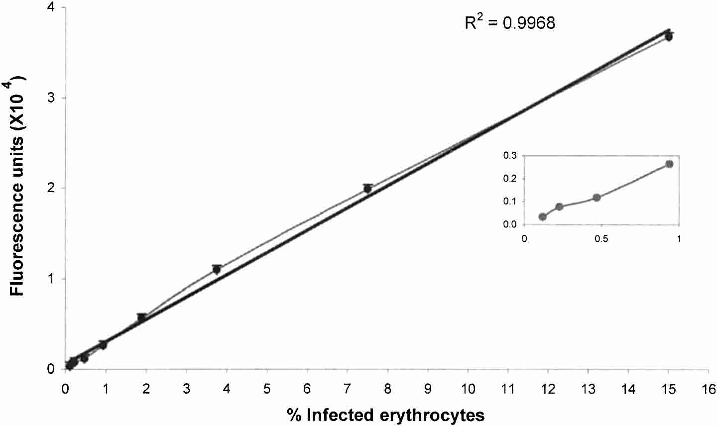
FIGURE 1. Comparison of the percentage of Plasmodium falciparum−infected erythrocytes determined by microscopic counting with fluo-
rescence intensity obtained from the microfluorimetric technique. A serial two-fold dilution of a synchronized infected culture (15.0% ring stage)
with noninfected erythrocytes was used. Bars indicate the standard deviation of the mean for four independently processed samples. The inset
shows the relationship below 1% of parasitemia.
rescence signal between 0.1% and 15% of ring stage infected
presence of infected erythrocytes. No differences were ob-
served when nonsynchronized or D-sorbitol-synchronized
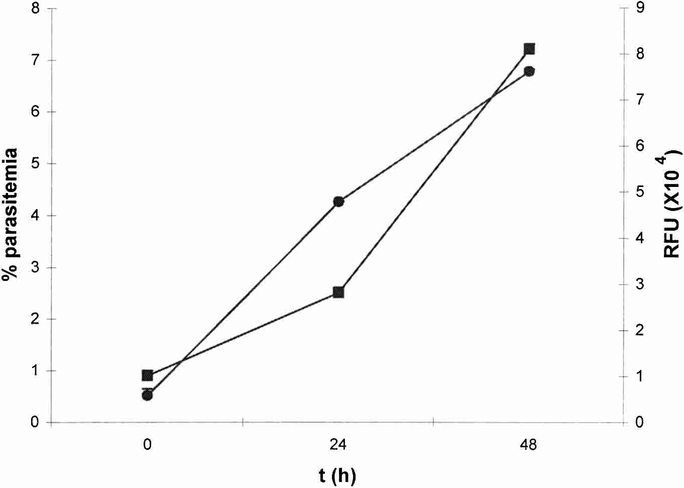
Time course for the assessment of parasitemia. Time Plasmodium cultures were used, nor were differences ob-
course experiments were then performed in which cultures of
served when chloroquine-sensitive (F32 and D6) or chloro-
P. falciparum- infected erythrocytes were initiated at a para-
quine-resistant (W2) strains were tested. Based upon these
sitemia of 0.5% and the number of parasites was determined
experiments, a time point of 48 hours was chosen for the
at different time intervals by both microscopic counting and
evaluation of potential anti-plasmodial compounds.
the microfluorimetric technique. Figure 2 shows that both
Determination of IC50 values of known antimalarial
methods of detection are equally effective in detecting the
drugs. The microfluorimetric method was used to determine
FIGURE 2. Time course experiments with Plasmodium falciparum−infected erythrocytes by microscopic counting and the microfluorimetric
techniques. Parallel cultures of synchronized parasites were initiated at a parasitemia of 0.5% and analyzed at 24 and 48 hours (h). Bars indicate
the standard deviation of the mean for two independently processed samples. RFU ס relative fluorescence units.
the effect of known antimalarial drugs on the growth of P.
its utility as a systematic and efficient means of screening
falciparum by testing the effect of chloroquine and meflo-
large numbers of crude extracts. We considered as active
quine on the growth on the F32 strain, a chloroquine-
those plant extracts with IC50 values < 50 g/mL. Table 1
susceptible parasite. From dose-response experiments, an
shows that there was a perfect correlation between the radio-
IC50 of 31 ± 0.7 nM (mean ± SD) for chloroquine was deter-
activity-based, microscopic, and microfluorimetric techniques
mined using the microfluorimetric method, which is compa-
with respect to their ability to detect plant extracts with anti-
rable to the previously reported value of 29 ± 9 nM deter-
plasmodial activity (seven of seven extracts tested with the
mined by 3H-hypoxanthine incorporation.15 The IC50 for me-
three assays and two of two extracts tested with the fluori-
floquine was 15 ± 3.7 nM, which is comparable to the value of
metric and radioactivity methods). While the IC50 levels of
9.2 ± 4.2 nM that was determined with the radioactivity-based
crude extracts measured by the radioactivity-based and mi-
method.14 The dose response curves obtained with the radio-
croscopic methods tend to be lower than those values mea-
activity-based and microfluorimetric methods for measuring
sure by the microfluorimetric assay, no differences were ob-
the effect of chloroquine on the growth of the chloroquine-
served in IC50 values when pure compounds were evaluated
resistant W2 clone are shown in Figure 3. We did not observe
(Figure 3). We carried out the complementary experiment in
any significant difference in the IC50 values determined by
which plants shown to be inactive by the radioactivity-based
either method, yielding IC50 values of 86.5 ± 9 and 88.7 ± 0.72
method were tested in the microfluorimetric assay. In every
nM for the radioactivity-based and microfluorimetric meth-
case (five of five), plants that were inactive in the radioactiv-
ods, respectively. The IC50 values determined for chloroquine
ity-based assay were also inactive in the microfluorimetric
in these experiments are comparable to the published value of
method, an observation relevant to the use of the latter
128 ± 73 nM for the chloroquine-resistant strains.5,15
method for drug discovery (Table 1). Drug discovery. Natural products from plants have been a
The microfluorimetric assay was used to guide the purifi-
rich source of anti-parasitic compounds.7,8 Therefore, we
cation of a compound with anti-Plasmodium activity from the
evaluated the ability of the microfluorimetric method to de-
plant Coccoloba parimensis. Initial screening of a crude ex-
tect plant extracts with anti-plasmodial activity and to assess
tract of leaves of C. parimensis demonstrated significant anti-
plasmodial activity (IC50 ס 6−12 g/mL). The extract was
subjected to liquid-liquid partition with hexane, ethyl acetate,
methanol and water, a technique used to separate the chemi-
cal constituents on the basis of their relative polarity12 and the
fractions were tested for anti-plasmodial activity. Purifica-
tion of the sample resultant from the ethyl acetate fraction
(IC50 ס 10 g/mL) led to the isolation of the methyl ester of
gallic acid that showed IC50 values < 2 g/mL.16
The microfluorimetric method for detecting anti-plas-
modial compounds described herein has several advantages
over the traditional assay that monitors the incorporation
of 3H-hypoxanthine by the parasite.3 The radioactivity-
based method requires the use of an expensive, hazardous
radioactive compound, costly liquid -scintillation counter
Comparison of IC50 values for crude plant extracts by uptake of
[3H]-hypoxanthine, microscopic counting of Giemsa-stained thin
blood smears, and the microfluorimetric technique*
FIGURE 3. Determination of the 50% inhibitory concentration
(IC50) values for chloroquine by the incorporation of 3H-
hypoxanthine (top) and the microfluorimetric technique (bottom).
Cultures of Plasmodium falciparum W2 strain-infected erythrocytes
were initiated at a parasitemia of 0.5%, incubated with different con-
centrations of chloroquine, and the number of parasites was deter-
mined at 48 hours. IC50 values of 88.7 and 86.5 g/mL were deter-
mined for the microfluorimetric and radioactivity-based assays, re-
spectively. Bars indicate the standard deviation from the mean for
four independently processed samples. CPM ס counts per minute;
* Values are in micrograms/milliliter.
IC50 ס 50% inhibitory concentration; ND ס not done.
FLUORIMETRIC METHOD FOR DETECTION OF ANTI-MALARIAL DRUGS
equipment, and special local regulations for the introduction,
Received April 4, 2003. Accepted for publication October 1, 2003.
management, and disposal of radioactive waste. An impedi-
Acknowledgments: Special thanks are given to Phil Rosenthal, Den-
ment for the development of drug discovery programs in de-
nis Kyle, and Jeff Ryan for their continuous and generous support.
veloping countries is the lack of accessible and appropriate
We also thank all members of the laboratory of Eduardo Ortega-
technology that would permit the efficient testing of biologic
Barria for helpful discussions and encouragement.
materials for anti-plasmodial activity. Although several non-
Financial support: This work was supported by the International Co-
radioactivity-based methods have been developed over the
operative Biodiversity Groups Program, award #1U01 TW01021-01.
The laboratory of Eduardo Ortega-Barria is partially supported by
years, they are cumbersome, multistep procedures.4,5
National Institutes of Health grant 1R03 TW01076.
The method described herein is based upon the detection
of Plasmodium DNA in short-term cultures using a 96-well
Authors’ addresses: Yolanda Corbett, Liuris Herrera, Jose Gonzalez,
Luz I. Romero, and Eduardo Ortega-Barría, Instituto de Investiga-
format, allowing the efficient and quantitative measurements
ciones Científicas Avanzadas y Servicios de Alta Tecnología, Ciudad
of anti-plasmodial activity in a large number of samples. The
del Saber, PO Box 7250, Zona 5, Clayton, Panama City, Panama. Luis
method uses PicoGreen®, an ultrasensitive fluorophore that
Cubilla, Laboratorio de Productos Naturales, Universidad de
intercalates into the double-stranded DNA of Plasmodium in
Panama, Panama City, Panama. Todd L. Capson, Smithsonian Tropi-
cal Research Institute, Apartado 2072, Balboa, Ancon, Panama.
solution, enabling the detection of as little as 25 pg/ml of
Phyllis D. Coley and Thomas Kursar, Department of Biology, Uni-
dsDNA, a 400-fold increase in sensitivity compared with the
versity of Utah, Salt Lake City, UT 84112 and Smithsonian Tropical
DNA intercalator Hoechst 33258 (Polysciences, Inc., War-
Research Institute, Apartado 2072, Balboa, Ancon, Panama.
Reprint requests: Eduardo Ortega-Barría, Instituto de Investiga-
The PicoGreen® method is straightforward and rapid. The
ciones Científicas Avanzadas y Servicios de Alta Tecnología, Ciudad
parasites are first incubated with the test drug for 48 hours,
del Saber, PO Box 7250, Zona 5, Clayton, Panama City, Panama, Tele-
phone: 507-317-0012, Fax: 507-317-0023, E-mail: eortega@senacyt.
followed by addition of PicoGreen®, followed by a 5−30-
minute incubation period prior to the measurement of fluo-rescence. The PicoGreen® assay protocol presented herein issimpler than that for Hoechst 33258 since there is no require-
ment to remove potentially interfering compounds such as
1. Greenwood B, Mutabingwa T, 2002. Malaria in 2002. Nature 415:
hemoglobin and hemozoin, nor is there a chloroform extrac-
tion step to prevent quenching of fluorescence.17 The repli-
2. Riddley RG, 1999. Planting the seeds of new antimalarial drugs.
cation of the parasite is directly proportional to the amount of
fluorescence, with a linear relationship between parasitemias
3. Desjardins RE, Canfield CJ, Haynes JD, Chulay JD, 1979. Quan-
titative assessment of antimalarial activity in vitro by a semi-
of 0.1% and 15%. We have used synchronized and non-
automated microdilution technique. Antimicrob Agents
synchronized parasites, and observed no significant differ-
ences. In addition, the samples can be stored at −20°C and
4. Makler MT, Gibbins BL, 1991. Laboratory diagnosis of malaria.
read at a more convenient time without a significant change in
the fluorescence signal. Significantly, if a fluorescence micro-
5. Delhaes L, Lazaro JE, Gay F, Thellier M, Danis M, 1999. The
microculture tetrazolium assay (MTA): another colorimetric
plate reader is not available, determination of parasite growth
method of testing Plasmodium falciparum chemosensitivity.
may be achieved with a less-expensive minifluorimeter (Mini-
Annals Trop Med Parasitol 93: 31–40.
fluorimeter TKO 100; Hoefer Scientific Instruments, San
6. Makler MT, Hinrichs DJ, 1993. Measurement of the lactate de-
hydrogenase activity of Plasmodium falciparum as an assess-
ment of parasitemia. Am J Trop Med Hyg 48: 205–210.
We compared the microfluorimetric methodology with the
7. Klayman DL, 1993. Artemisia annua, from weed to respectable
conventional radioactivity-based assay by using both methods
antimalarial plant. Kinghorn AD, Balandron MA, eds. Human
to test crude plant extracts for anti-plasmodial activity. We
Medicinal Agents from Plants. Washington, DC: American
found that for all of the extracts tested, both methods yielded
identical results. We do not have an explanation for the small
8. Muñoz V, Sauvain M, Bourdy G, Callapa J, Bergeron S, Rojas I,
Bravo JA, Balderrama L, Ortiz B, Gimenez A, DeHaro E,
differences between the calculated IC50 values of crude plant
2000. A search for natural bioactive compounds in Bolivia
extracts as determined by the two methods. One possible
through a multidisciplinary approach Part I. Evaluation of the
explanation is the presence of low levels of interfering sub-
antimalarial activity of plants used by the Chacobo Indians. J
stances in the extracts. Alternatively, the persistence of Plas-Ethnopharmacol 69: 127–137. modium-derived DNA related to the initial parasite inoculum
9. Kursar TA, Capson TL, Coley PD, Corley DG, Gupta MB, Har-
rison LA, Ortega-Barría E, Windsor DM, 1999. Ecologically
may be responsible. However, no significant difference in
guided bioprospecting in Panama. Pharmaceut Biol 37 (suppl):
IC50 values were observed between the two methods when
pure compounds (chloroquine and mefloquine) were tested,
10. Trager W, Jensen JB, 1976. Human malaria parasites in continu-
supporting the utility the PicoGreen® assay for quantifying
ous culture. Science 193: 673–675.
anti-plasmodial activity. The microfluorimetric method de-
11. Lambros C, Vanderberg JP, 1979. Synchronization of Plasmo-dium falciparum erythrocytic stages in culture. J Parasitol 65:
scribed herein has been used successfully to guide the purifi-
cation of compounds with anti-plasmodial activity from crude
12. Montenegro H, Gutiérrez M, Romero LI, Ortega-Barría E, Cap-
plant extracts. It is hoped that the development of an effec-
son TL, Cubilla-Rios L, 2003. Aporphine alkaloids from Guat-
tive and straightforward method for measuring anti-
teria spp. with leishmanicidal activity. Planta Med 69: 677–679.
plasmodial activity that does not use radioactive isotopes will
13. DeHaro E, Gautret P, Munoz V, Sauvain M, 2000. Evaluación de
stimulate anti-malarial drug discovery programs in a number
la actividad antimalarica in vitro de productos naturales o de
sintesis. Técnicas de Laboratorio para la Selección de Sustan-
of countries, in particular, those most affected by this deadly
cias Antimalaricas. CYTED. La Paz, Bolivia: Imprenta Perez,
14. Basco LK, Marquet F, Makler MT, Le Bras J, 1995. Plasmodium
16. Westenburg HE, Lee KJ, Lee SK, Fong HHS, Van Breemen RB,
falciparum and Plasmodium vivax: Lactate dehydrogenase ac-
Pezzuto JM, Kinghorn DA, 2000. Activity-guided isolation of
tivity and its application for in vitro drug susceptibility assay.
antioxidative constituents of Cotinus coggygria. J Nat Prod 63:
15. Makler MT, Ries JM, Williams JA, Bancroft JE, Piper RC, Gib-
17. Smeijsters LJJW, Zijlstra NM, Franssen FFJ, Overdule JP, 1996.
bins BL, Hinrichs DJ, 1993. Parasite lactate dehydrogenase as
Simple, fast, and accurate fluorimetric method to determine
an assay for Plasmodium falciparum drug sensitivity. Am J
drug susceptibility of Plasmodium falciparum in 24-well sus-
pension cultures. Antimicrobial Agents Chemother 40: 835–838.
PPTP y L2TP por Marisabel Rodriguez Bilardo Los protocolos PPTP y L2TP nacieron para crear VPNs. Además de permitir crear túneles a través de Internet, pueden encriptar los datos enviados y autenticar a los usuarios. Las VPNs conectan sitios remotos en forma segura y ademásbajan costos porque utilizan redes públicas en vez de líneasPPTP y L2TP son idénticos en la c
VAD Congress 2014: June 11th – June 14th in Bayreuth Panel: Afrikanische Kapitalismen ABSTRACT Economic and Cultural Aspects of Local Capitalisms: Milieus, Practices and Life Styles in the Middle-Classes of Nairobi and Mombasa Bayreuth Academy of Advanced African Studies / Development Sociology, University of Bayreuth This paper wants to test the notion of capitalism on local p
 FLUORIMETRIC METHOD FOR DETECTION OF ANTI-MALARIAL DRUGS
FIGURE 1. Comparison of the percentage of Plasmodium falciparum−infected erythrocytes determined by microscopic counting with fluo-
rescence intensity obtained from the microfluorimetric technique. A serial two-fold dilution of a synchronized infected culture (15.0% ring stage)
with noninfected erythrocytes was used. Bars indicate the standard deviation of the mean for four independently processed samples. The inset
FLUORIMETRIC METHOD FOR DETECTION OF ANTI-MALARIAL DRUGS
FIGURE 1. Comparison of the percentage of Plasmodium falciparum−infected erythrocytes determined by microscopic counting with fluo-
rescence intensity obtained from the microfluorimetric technique. A serial two-fold dilution of a synchronized infected culture (15.0% ring stage)
with noninfected erythrocytes was used. Bars indicate the standard deviation of the mean for four independently processed samples. The inset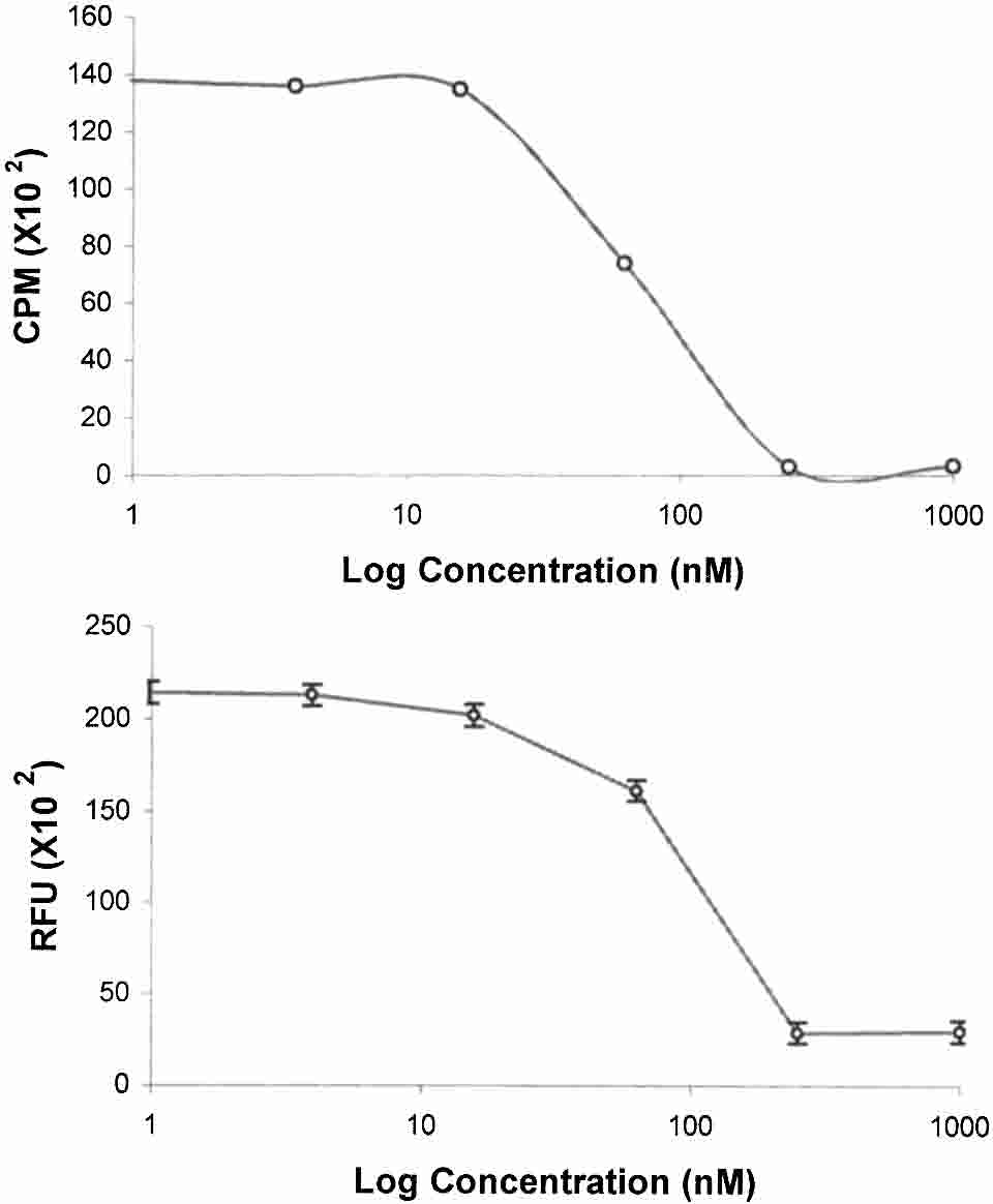 the effect of known antimalarial drugs on the growth of P.
its utility as a systematic and efficient means of screening
falciparum by testing the effect of chloroquine and meflo-
large numbers of crude extracts. We considered as active
quine on the growth on the F32 strain, a chloroquine-
those plant extracts with IC50 values < 50 g/mL. Table 1
susceptible parasite. From dose-response experiments, an
shows that there was a perfect correlation between the radio-
IC50 of 31 ± 0.7 nM (mean ± SD) for chloroquine was deter-
activity-based, microscopic, and microfluorimetric techniques
mined using the microfluorimetric method, which is compa-
with respect to their ability to detect plant extracts with anti-
rable to the previously reported value of 29 ± 9 nM deter-
plasmodial activity (seven of seven extracts tested with the
mined by 3H-hypoxanthine incorporation.15 The IC50 for me-
three assays and two of two extracts tested with the fluori-
floquine was 15 ± 3.7 nM, which is comparable to the value of
metric and radioactivity methods). While the IC50 levels of
9.2 ± 4.2 nM that was determined with the radioactivity-based
crude extracts measured by the radioactivity-based and mi-
method.14 The dose response curves obtained with the radio-
croscopic methods tend to be lower than those values mea-
activity-based and microfluorimetric methods for measuring
sure by the microfluorimetric assay, no differences were ob-
the effect of chloroquine on the growth of the chloroquine-
served in IC50 values when pure compounds were evaluated
resistant W2 clone are shown in Figure 3. We did not observe
(Figure 3). We carried out the complementary experiment in
any significant difference in the IC50 values determined by
which plants shown to be inactive by the radioactivity-based
either method, yielding IC50 values of 86.5 ± 9 and 88.7 ± 0.72
method were tested in the microfluorimetric assay. In every
nM for the radioactivity-based and microfluorimetric meth-
case (five of five), plants that were inactive in the radioactiv-
ods, respectively. The IC50 values determined for chloroquine
ity-based assay were also inactive in the microfluorimetric
in these experiments are comparable to the published value of
method, an observation relevant to the use of the latter
128 ± 73 nM for the chloroquine-resistant strains.5,15
method for drug discovery (Table 1).
the effect of known antimalarial drugs on the growth of P.
its utility as a systematic and efficient means of screening
falciparum by testing the effect of chloroquine and meflo-
large numbers of crude extracts. We considered as active
quine on the growth on the F32 strain, a chloroquine-
those plant extracts with IC50 values < 50 g/mL. Table 1
susceptible parasite. From dose-response experiments, an
shows that there was a perfect correlation between the radio-
IC50 of 31 ± 0.7 nM (mean ± SD) for chloroquine was deter-
activity-based, microscopic, and microfluorimetric techniques
mined using the microfluorimetric method, which is compa-
with respect to their ability to detect plant extracts with anti-
rable to the previously reported value of 29 ± 9 nM deter-
plasmodial activity (seven of seven extracts tested with the
mined by 3H-hypoxanthine incorporation.15 The IC50 for me-
three assays and two of two extracts tested with the fluori-
floquine was 15 ± 3.7 nM, which is comparable to the value of
metric and radioactivity methods). While the IC50 levels of
9.2 ± 4.2 nM that was determined with the radioactivity-based
crude extracts measured by the radioactivity-based and mi-
method.14 The dose response curves obtained with the radio-
croscopic methods tend to be lower than those values mea-
activity-based and microfluorimetric methods for measuring
sure by the microfluorimetric assay, no differences were ob-
the effect of chloroquine on the growth of the chloroquine-
served in IC50 values when pure compounds were evaluated
resistant W2 clone are shown in Figure 3. We did not observe
(Figure 3). We carried out the complementary experiment in
any significant difference in the IC50 values determined by
which plants shown to be inactive by the radioactivity-based
either method, yielding IC50 values of 86.5 ± 9 and 88.7 ± 0.72
method were tested in the microfluorimetric assay. In every
nM for the radioactivity-based and microfluorimetric meth-
case (five of five), plants that were inactive in the radioactiv-
ods, respectively. The IC50 values determined for chloroquine
ity-based assay were also inactive in the microfluorimetric
in these experiments are comparable to the published value of
method, an observation relevant to the use of the latter
128 ± 73 nM for the chloroquine-resistant strains.5,15
method for drug discovery (Table 1).